OslerThoracic Key Points on Lung Cancer:
- Non-small cell lung cancer is a disease in which malignant (cancer) cells form in the tissues of the lung.There are several types of non-small cell lung cancer. The two most common types of lung cancer are Small Cell and Non-Small Cell lung cancer
- Smoking is the major risk factor for non-small cell lung cancer. Other risk factors include asbestos exposure and radon exposure.
- Signs of non-small cell lung cancer include a cough that doesn't go away and shortness of breath. Signs of more advanced cancer include hoarseness, severe headaches, new bone pain
- Your physician will order tests that examine the lungs and surrounding areas. They are used to diagnose, and stage non-small cell lung cancer.
- Certain factors affect prognosis (chance of recovery) and treatment options.
- Small Cell lung cancer is usually treated with chemo-therapy and radiation therapy. Surgery is infrequently used.
- Non-small cell lung cancer treatment depends on stage. Stage 1 and 2 lung cancer is frequently treated with surgery, whereas stage 3 and 4 is most often treated with chemotherapy and/or radiation therapy.
What is Lung Cancer?
Understanding the Lungs & Non-Small Cell Lung Cancer
The lungs are a pair of cone-shaped breathing organs in the chest. The lungs bring oxygen into the body as you breathe in. They release carbon dioxide, a waste product of the body’s cells, as you breathe out. Each lung has sections called lobes. The left lung has two lobes. The right lung is slightly larger and has three lobes. Two tubes called bronchi lead from the trachea (windpipe) to the right and left lungs. The bronchi are sometimes also involved in lung cancer. Tiny air sacs called alveoli and small tubes called bronchioles make up the inside of the lungs.
A thin membrane called the pleura covers the outside of each lung and lines the inside wall of the chest cavity. This creates a sac called the pleural cavity. The pleural cavity normally contains a small amount of fluid that helps the lungs move smoothly in the chest when you breathe.
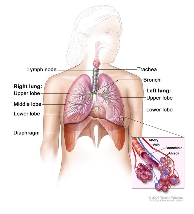
Figure 1:Anatomy of the respiratory system, showing the trachea and both lungs and their lobes and airways. Lymph nodes and the diaphragm are also shown. Oxygen is inhaled into the lungs and passes through the thin membranes of the alveoli and into the bloodstream (see inset).
There are two main types of lung cancer: non-small cell lung cancer and small cell lung cancer.
There are several types of non-small cell lung cancer.
Each type of non-small cell lung cancer has different kinds of cancer cells. The cancer cells of each type grow and spread in different ways. The types of non-small cell lung cancer are named for the kinds of cells found in the cancer and how the cells look under a microscope:
- Squamous cell carcinoma: Cancer that begins in squamous cells, which are thin, flat cells that look like fish scales. This is also called epidermoid carcinoma.
- Adenocarcinoma: Cancer that begins in the cells that line the alveoli and make substances such as mucus.
- Large cell carcinoma: Cancer that may begin in several types of large cells.
- Other less common types of non-small cell lung cancer are: pleomorphic, carcinoid tumor, salivary gland carcinoma, and unclassified carcinoma
What Are the Risk Factors for Lung Cancer?
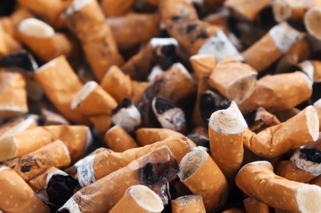
Smoking is the major risk factor for non-small cell lung cancer.
Anything that increases your chance of getting a disease is called a risk factor. Having a risk factor does not mean that you will get cancer; not having risk factors doesn't mean that you will not get cancer. Talk to your doctor if you think you may be at risk for lung cancer.
Risk factors for lung cancer include the following:
- Smoking cigarettes, pipes, or cigars, now or in the past. This is the most important risk factor for lung cancer. The earlier in life a person starts smoking, the more often a person smokes, and the more years a person smokes, the greater the risk of lung cancer.
- Being exposed to secondhand smoke.
-
- Being exposed to radiation from any of the following:
- - Radiation therapy to the breast or chest.
- - Radon in the home or workplace.
- - Imaging tests such as CT scans.
- Being exposed to asbestos, chromium, nickel, beryllium, arsenic, soot, or tar in the workplace.
- Living where there is air pollution.
- Having a family history of lung cancer.
- Older age is the main risk factor for most cancers.
The chance of getting cancer increases as you get older.
When smoking is combined with other risk factors, the risk of lung cancer is increased.
What Are the Symptoms?
Signs of non-small cell lung cancer include a cough that doesn't go away and shortness of breath.
Sometimes lung cancer does not cause any signs or symptoms. It may be found during a chest x-ray done for another condition. Signs and symptoms may be caused by lung cancer or by other conditions. Check with your doctor if you have any of the following:
- Chest discomfort or pain.
- A cough that doesn’t go away or gets worse over time.
- Trouble breathing.
- Wheezing.
- Blood in sputum (mucus coughed up from the lungs).
- Hoarseness.
- Loss of appetite.
- Weight loss for no known reason.
- Feeling very tired.
- Trouble swallowing.
- Swelling in the face and/or veins in the neck.
Survival With Lung Cancer
Certain factors affect prognosis (chance of recovery) and treatment options.
The prognosis (chance of recovery) and treatment options depend on the following:
- The stage of the cancer (the size of the tumor and whether it is in the lung only or has spread to other places in the body).
- The type of lung cancer.
- Whether the cancer has mutations (changes) in certain genes, such as the epidermal growth factor receptor (EGFR) gene or the anaplastic lymphoma kinase (ALK) gene.
- Whether there are signs and symptoms such as coughing or trouble breathing.
- The patient’s general health.
Minimally Invasive Lobectomy
this section includes description of how procedure is done
Before Your Surgery (Pre-Op)
Tests Before Your Surgery
Tests that examine the lungs are used to detect (find), diagnose, and stage non-small cell lung cancer.
Tests and procedures to detect, diagnose, and stage non-small cell lung cancer are often done at the same time. Some of the following tests and procedures may be used:
Physical exam and history
An exam of the body to check general signs of health, including checking for signs of disease, such as lumps or anything else that seems unusual. A history of the patient’s health habits, including smoking, and past jobs, illnesses, and treatments will also be taken.
Laboratory tests
Medical procedures that test samples of tissue, blood, urine, or other substances in the body. These tests help to diagnose disease, plan and check treatment, or monitor the disease over time.
Chest x-ray
An x-ray of the organs and bones inside the chest. An x-ray is a type of energy beam that can go through the body and onto film, making a picture of areas inside the body.
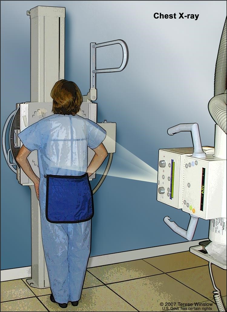
Figure 2: X-ray of the chest. X-rays are used to take pictures of organs and bones of the chest. X-rays pass through the patient onto film.
CT scan (CAT scan)
A procedure that makes a series of detailed pictures of areas inside the body, such as the chest, taken from different angles. The pictures are made by a computer linked to an x-ray machine. A dye may be injected into a vein or swallowed to help the organs or tissues show up more clearly. This procedure is also called computed tomography, computerized tomography, or computerized axial tomography.
Sputum cytology
A procedure in which a pathologist views a sample of sputum (mucus coughed up from the lungs) under a microscope, to check for cancer cells.
Fine-needle aspiration (FNA) biopsy of the lung
The removal of tissue or fluid from the lung using a thin needle. A CT scan, ultrasound, or other imaging procedure is used to locate the abnormal tissue or fluid in the lung. A small incision may be made in the skin where the biopsy needle is inserted into the abnormal tissue or fluid. A sample is removed with the needle and sent to the laboratory. A pathologist then views the sample under a microscope to look for cancer cells. A chest x-ray is done after the procedure to make sure no air is leaking from the lung into the chest.
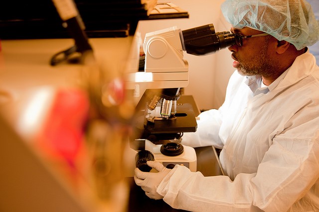
Figure 3: Dr. Ismael from William Osler Health System reviews a lung biopsy under the microscope
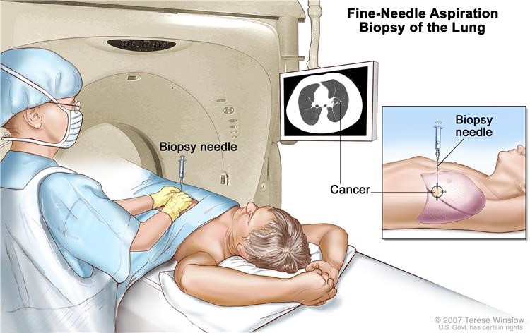
Figure 4: Fine-needle aspiration biopsy of the lung. The patient lies on a table that slides through the computed
tomography (CT) machine, which takes x-ray pictures of the inside of the body. The x-ray pictures help the doctor see where the abnormal tissue is in the lung. A biopsy needle is inserted through the chest wall and into the area of abnormal lung tissue. A small piece of tissue is removed through the needle and checked under the microscope for signs of cancer.
Bronchoscopy
A procedure to look inside the trachea and large airways in the lung for abnormal areas. A bronchoscope is inserted through the nose or mouth into the trachea and lungs. A bronchoscope is a thin, tube-like instrument with a light and a lens for viewing. It may also have a tool to remove tissue samples, which are checked under a microscope for signs of cancer.
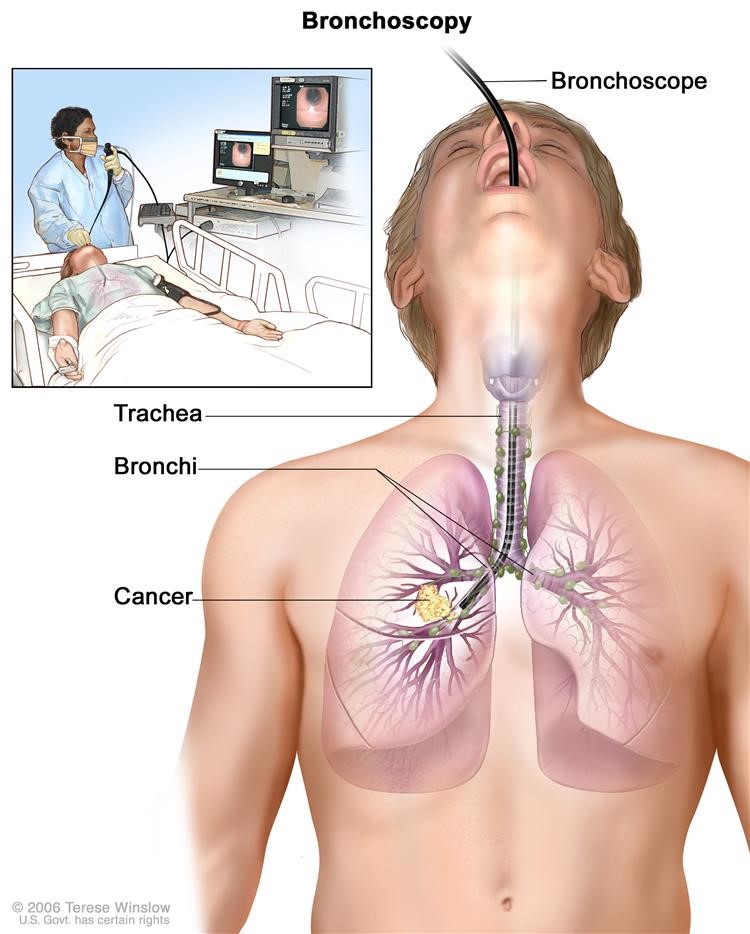
Figure 5: A bronchoscope is inserted through the mouth, trachea, and major bronchi into the lung, to look for abnormal areas. A bronchoscope is a thin, tube-like instrument with a light and a lens for viewing. It may also have a cutting tool. Tissue samples may be taken to be checked under a microscope for signs of disease.
Thoracentesis
The removal of fluid from the space between the lining of the chest and the lung, using a needle. A pathologist views the fluid under a microscope to look for cancer cells.
Light and electron microscopy
A laboratory test in which cells in a sample of tissue are viewed under regular and high-powered microscopes to look for certain changes in the cells.
Immunohistochemistry
A test that uses antibodies to check for certain antigens in a sample of tissue. The antibody is usually linked to a radioactive substance or a dye that causes the tissue to light up under a microscope. This type of test may be used to tell the difference between different types of cancer.
Staging Lung Cancer: Stages of Non-Small Cell Lung Cancer
KEY POINTS
After lung cancer has been diagnosed, tests are done to find out if cancer cells have spread within the lungs or to other parts of the body.
The following stages are used for non-small cell lung cancer:
- Occult (hidden) stage
- Stage 0 (carcinoma in situ)
- Stage I
- Stage II
- Stage IIIA
- Stage IIIB
- Stage IV
After lung cancer has been diagnosed, tests are done to find out if cancer cells have spread within the lungs or to other parts of the body.
The process used to find out if cancer has spread within the lungs or to other parts of the body is called staging. The information gathered from the staging process determines the stage of the disease. It is important to know the stage in order to plan treatment. Some of the tests used to diagnose non-small cell lung cancer are also used to stage the disease.
Other tests and procedures that may be used in the staging process include the following:
MRI (magnetic resonance imaging)
A procedure that uses a magnet, radio waves, and a computer to make a series of detailed pictures of areas inside the body, such as the brain. This procedure is also called nuclear magnetic resonance imaging (NMRI).
PET scan (positron emission tomography scan)
A procedure to find malignant tumor cells in the body. A small amount of radioactive glucose (sugar) is injected into a vein. The PET scanner rotates around the body and makes a picture of where glucose is being used in the body. Malignant tumor cells show up brighter in the picture because they are more active and take up more glucose than normal cells do.
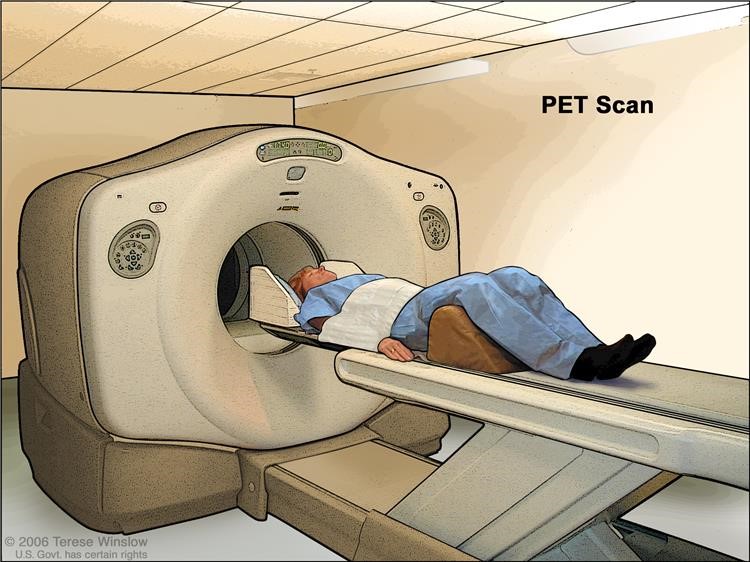 Figure 6: PET (positron emission tomography) scan. The patient lies on a table that slides through the PET machine. The head rest and white strap help the patient lie still. A small amount of radioactive glucose (sugar) is injected into the patient's vein, and a scanner makes a picture of where the glucose is being used in the body. Cancer cells show up brighter in the picture because they take up more glucose than normal cells do.
Radionuclide bone scan
A procedure to check if there are rapidly dividing cells, such as cancer cells, in the bone. A very small amount of radioactive material is injected into a vein and travels through the bloodstream. The radioactive material collects in the bones and is detected by a scanner.
Endoscopic ultrasound (EUS)
A procedure in which an endoscope is inserted into the body. An endoscope is a thin, tube-like instrument with a light and a lens for viewing. A probe at the end of the endoscope is used to bounce high-energy sound waves (ultrasound) off internal tissues or organs and make echoes. The echoes form a picture of body tissues called a sonogram. This procedure is also called endosonography. EUS may be used to guide fine needle aspiration (FNA) biopsy of the lung, lymph nodes, or other areas.
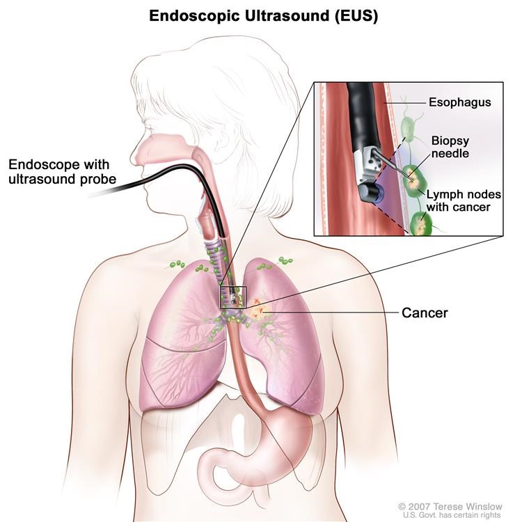
Figure 7: Endoscopic ultrasound-guided fine-needle aspiration biopsy: An endoscope that has an ultrasound probe and a biopsy needle is inserted through the mouth and into the esophagus. The probe bounces sound waves off body tissues to make echoes that form a sonogram (computer picture) of the lymph nodes near the esophagus. The sonogram helps the doctor see where to place the biopsy needle to remove tissue from the lymph nodes. This tissue is checked under a microscope for signs of cancer.
Mediastinoscopy
A surgical procedure to look at the organs, tissues, and lymph nodes between the lungs for abnormal areas. An incision (cut) is made at the top of the breastbone and a mediastinoscope is inserted into the chest. A mediastinoscope is a thin, tube-like instrument with a light and a lens for viewing. It may also have a tool to remove tissue or lymph node samples, which are checked
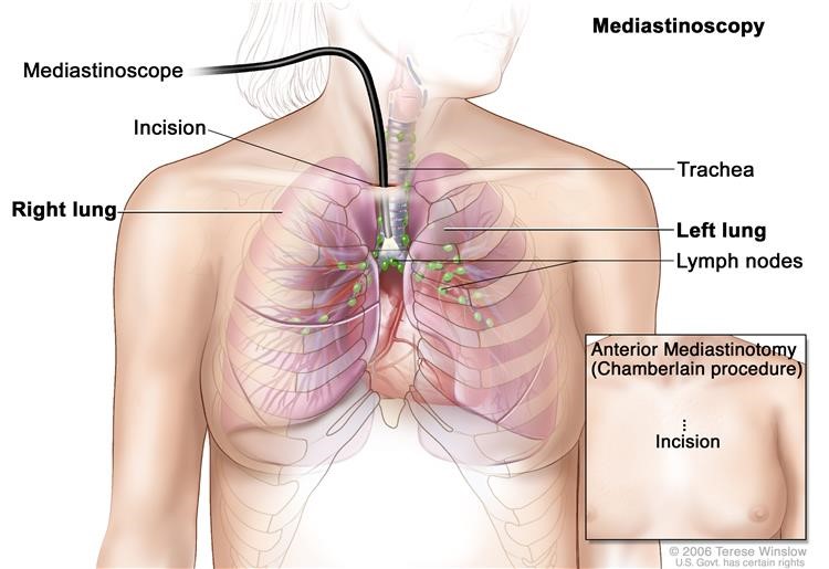
Figure 8: Mediastinoscopy. A mediastinoscope is inserted into the chest through an incision above the breastbone to look for abnormal areas between the lungs. A mediastinoscope is a thin, tube-like instrument with a light and a lens for viewing. It may also have a cutting tool. Tissue samples may be taken from lymph nodes on the right side of the chest and checked under a microscope for signs of cancer. In an anterior mediastinotomy (Chamberlain procedure), the incision is made beside the breastbone to remove tissue samples from the lymph nodes on the left side of the chest.
Lymph node biopsy
The removal of all or part of a lymph node. A pathologist views the tissue under a microscope to look for cancer cells.
Bone marrow aspiration and biopsy
The removal of bone marrow, blood, and a small piece of bone by inserting a hollow needle into the hipbone or breastbone. A pathologist views the bone marrow, blood, and bone under a microscope to look for signs of cancer.
There are three ways that cancer spreads in the body.
Cancer can spread through tissue, the lymph system, and the blood:
Tissue. The cancer spreads from where it began by growing into nearby areas.
Lymph system. The cancer spreads from where it began by getting into the lymph system. The cancer travels through the lymph vessels to other parts of the body.
Blood. The cancer spreads from where it began by getting into the blood. The cancer travels through the blood vessels to other parts of the body.
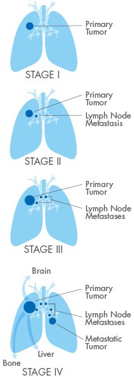
Cancer may spread from where it began to other parts of the body.
When cancer spreads to another part of the body, it is called metastasis. Cancer cells break away from where they began (the primary tumor) and travel through the lymph system or blood.
Lymph system. The cancer gets into the lymph system, travels through the lymph vessels, and forms a tumor (metastatic tumor) in another part of the body.
Blood. The cancer gets into the blood, travels through the blood vessels, and forms a tumor (metastatic tumor) in another part of the body.
The metastatic tumor is the same type of cancer as the primary tumor. For example, if non-small cell lung cancer spreads to the brain, the cancer cells in the brain are actually lung cancer cells. The disease is metastatic lung cancer, not brain cancer.
The following stages are used for non-small cell lung cancer:
Occult (hidden) stage
In the occult (hidden) stage, cancer cannot be seen by imaging or bronchoscopy. Cancer cells are found in sputum (mucus coughed up from the lungs) or bronchial washing (a sample of cells taken from inside the airways that lead to the lung). Cancer may have spread to other parts of the body.
Stage 0 (carcinoma in situ)
In stage 0, abnormal cells are found in the lining of the airways. These abnormal cells may become cancer and spread into nearby normal tissue. Stage 0 is also called carcinoma in situ.
Stage I
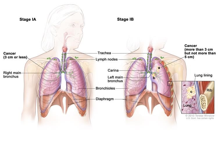
Figure 9: Stage IA and IB non-small cell lung cancer.
In stage I, cancer has formed. Stage I is divided into stages IA and IB:
Stage IA: The tumor is in the lung only and is 3 centimeters or smaller.
Stage IB: Cancer has not spread to the lymph nodes and one or more of the following is true:
- The tumor is larger than 3 centimeters but not larger than 5 centimeters.
- Cancer has spread to the main bronchus and is at least 2 centimeters below where the trachea joins the bronchus.
- Cancer has spread to the innermost layer of the membrane that covers the lung.
- Part of the lung has collapsed or developed pneumonitis (inflammation of the lung) in the area where the trachea joins the bronchus.
Stage II
Stage II is divided into stages IIA and IIB. Stage IIA and IIB are each divided into two sections depending on the size of the tumor, where the tumor is found, and whether there is cancer in the lymph nodes.
Stage IIA: Cancer has spread to certain lymph nodes on the same side of the chest as the primary tumor; the cancer is (a) 5 cm or smaller, (b) has spread to the main bronchus, and/or (c) has spread to the innermost layer of the lung lining. OR, cancer has not spread to lymph nodes; the cancer is (d) larger than 5 cm but not larger than 7 cm, (e) has spread to the main bronchus, and/or (f) has spread to the innermost layer of the lung lining. Part of the lung may have collapsed or become inflamed (not shown).
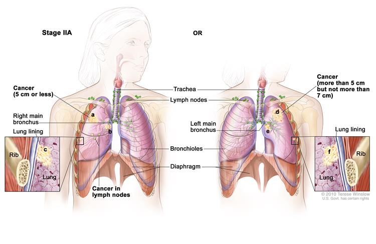
Figure 10: Stage IIA non-small cell lung cancer.
(1) Cancer has spread to lymph nodes on the same side of the chest as the tumor. The lymph nodes with cancer are within the lung or near the bronchus. Also, one or more of the following is true:
- The tumor is not larger than 5 centimeters.
- Cancer has spread to the main bronchus and is at least 2 centimeters below where the trachea joins the bronchus.
- Cancer has spread to the innermost layer of the membrane that covers the lung.
- Part of the lung has collapsed or developed pneumonitis (inflammation of the lung) in the area where the trachea joins the bronchus.
or (2) Cancer has not spread to lymph nodes and one or more of the following is true:
- The tumor is larger than 5 centimeters but not larger than 7 centimeters.
- Cancer has spread to the main bronchus and is at least 2 centimeters below where the trachea joins the bronchus.
- Cancer has spread to the innermost layer of the membrane that covers the lung.
- Part of the lung has collapsed or developed pneumonitis (inflammation of the lung) in the area where the trachea joins the bronchus.
Stage IIB:
Cancer has spread to certain lymph nodes on the same side of the chest as the primary tumor; the cancer is (a) larger than 5 cm but not larger than 7 cm, (b) has spread to the main bronchus, and/or (c) has spread to the innermost layer of the lung lining. Part of the lung may have collapsed or become inflamed (not shown). OR, (d) the cancer is larger than 7 cm; (e) has spread to the main bronchus, (f) the diaphragm, (g) the chest wall or the lining of the chest wall; and/or (h) has spread to the membrane around the heart. There may be one or more separate tumors in the same lobe of the lung; cancer may have spread to the nerve that controls the diaphragm; the whole lung may have collapsed or become inflamed (not shown).
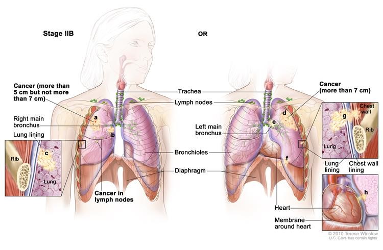
Figure 11: Stage IIB non-small cell lung cancer.
(1) Cancer has spread to nearby lymph nodes on the same side of the chest as the tumor. The lymph nodes with cancer are within the lung or near the bronchus. Also, one or more of the following is true:
- The tumor is larger than 5 centimeters but not larger than 7 centimeters.
- Cancer has spread to the main bronchus and is at least 2 centimeters below where the trachea joins the bronchus.
- Cancer has spread to the innermost layer of the membrane that covers the lung.
- Part of the lung has collapsed or developed pneumonitis (inflammation of the lung) in the area where the trachea joins the bronchus.
or(2) Cancer has not spread to lymph nodes and one or more of the following is true:
- The tumor is larger than 7 centimeters.
- Cancer has spread to the main bronchus (and is less than 2 centimeters below where the trachea joins the bronchus), the chest wall, the diaphragm, or the nerve that controls the diaphragm.
- Cancer has spread to the membrane around the heart or lining the chest wall.
- The whole lung has collapsed or developed pneumonitis (inflammation of the lung).
- There are one or more separate tumors in the same lobe of the lung.
Stage IIIA
Stage IIIA is divided into three sections depending on the size of the tumor, where the tumor is found, and which lymph nodes have cancer (if any).
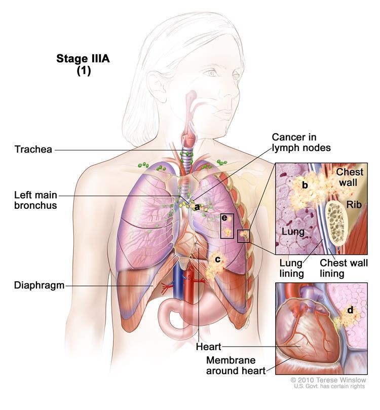
Figure 12: Stage IIIA non-small cell lung cancer.
(1) Cancer has spread to certain lymph nodes on the same side of the chest as the primary tumor. The cancer may have spread to (a) the main bronchus; (b) lung lining, chest wall lining, or chest wall; (c) diaphragm; and/or (d) membrane around the heart; and/or (e) there may be one or more separate tumors in the same lobe of the lung. Cancer may have spread to the nerve that controls the diaphragm, and part or all of the lung may have collapsed or become inflamed (not shown).
Cancer has spread to lymph nodes on the same side of the chest as the tumor. The lymph nodes with cancer are near the sternum (chest bone) or where the bronchus enters the lung. Also:
- The tumor may be any size.
- Part of the lung (where the trachea joins the bronchus) or the whole lung may have collapsed or developed pneumonitis (inflammation of the lung).
- There may be one or more separate tumors in the same lobe of the lung.
- Cancer may have spread to any of the following:
- Main bronchus, but not the area where the trachea joins the bronchus.
- Chest wall.
- Diaphragm and the nerve that controls it.
- Membrane around the lung or lining the chest wall.
- Membrane around the heart.
Or(2) Cancer has spread to certain lymph nodes on the same side of the chest as the primary tumor. The cancer may have spread to (a) the main bronchus; (b) the lung lining, chest wall lining, or chest wall; (c) diaphragm; (d) heart and/or membrane around the it; (e) major blood vessels that lead to or from the heart; (f) trachea; (g) esophagus; (h) sternum; and/or (i) carina; and/or (j) there may be one or more separate tumors in any lobe of the same lung. Cancer may have spread to the nerves that control the diaphragm and larynx, and the whole lung may have collapsed or become inflamed (not shown).
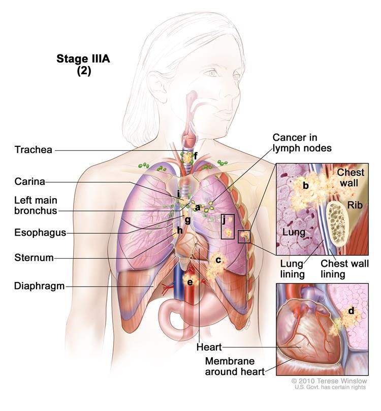
Figure 13: Stage IIIA lung cancer.
Cancer has spread to lymph nodes on the same side of the chest as the tumor. The lymph nodes with cancer are within the lung or near the bronchus. Also:
- The tumor may be any size.
- The whole lung may have collapsed or developed pneumonitis (inflammation of the lung).
- There may be one or more separate tumors in any of the lobes of the lung with cancer.
- Cancer may have spread to any of the following:
- Main bronchus, but not the area where the trachea joins the bronchus.
- Chest wall.
- Diaphragm and the nerve that controls it.
- Membrane around the lung or lining the chest wall.
- Heart or the membrane around it.
- Major blood vessels that lead to or from the heart.
- Trachea.
- Esophagus.
- Nerve that controls the larynx (voice box).
- Sternum (chest bone) or backbone.
- Carina (where the trachea joins the bronchi).
Or(3) Cancer has spread to (a) the heart; (b) major blood vessels that lead to or from the heart; (c) trachea; (d) esophagus; (e) sternum; and/or (f) carina. Cancer may have spread to the nerve that controls the larynx (not shown).
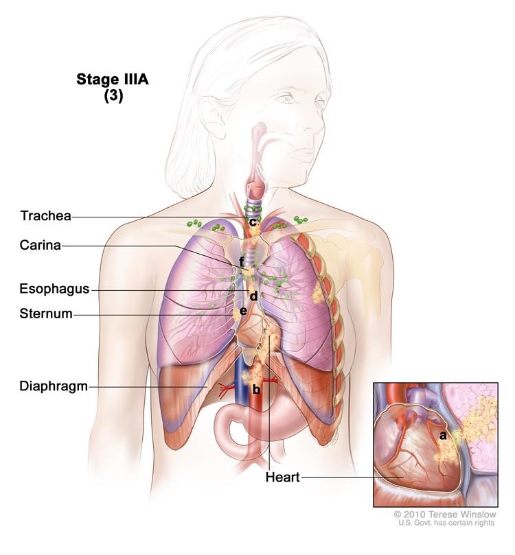
Figure 14: Stage IIIA non-small cell lung cancer.
Cancer has not spread to the lymph nodes and the tumor may be any size. Cancer has spread to any of the following:
- Heart.
- Major blood vessels that lead to or from the heart.
- Trachea.
- Esophagus.
- Nerve that controls the larynx (voice box).
- Sternum (chest bone) or backbone.
- Carina (where the trachea joins the bronchi).
Stage IIIB
Stage IIIB is divided into two sections depending on the size of the tumor, where the tumor is found, and which lymph nodes have cancer.
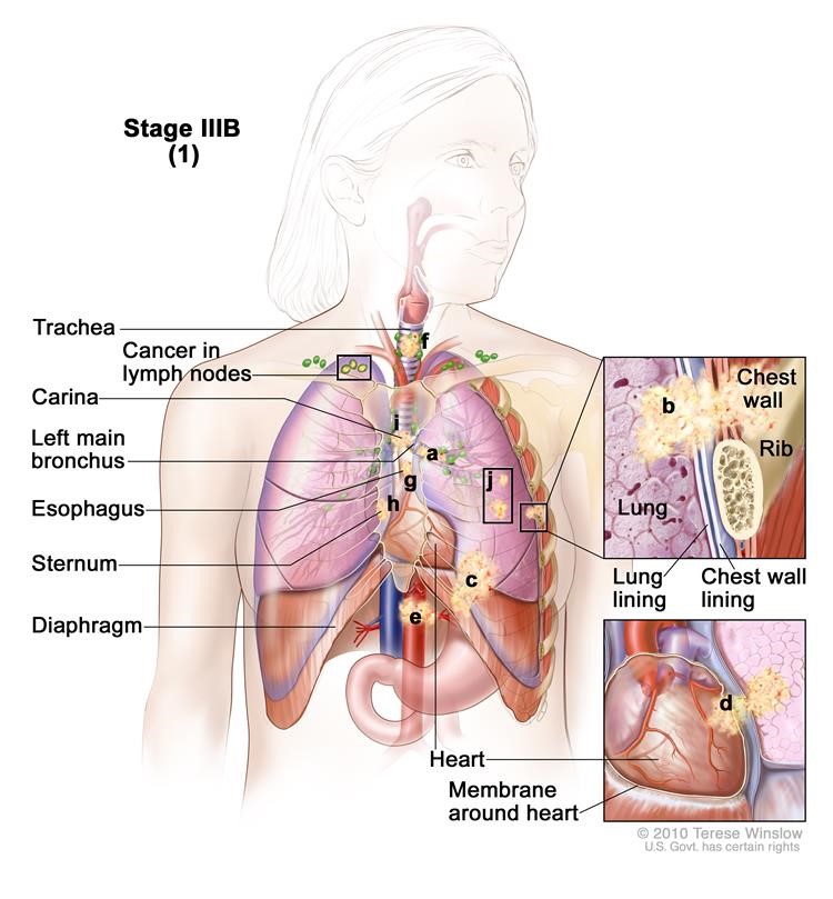
Figure 15: Stage IIIB non-small cell lung cancer.
(1) Cancer has spread to lymph nodes above the collarbone or on the opposite side of the chest as the primary tumor. The cancer may have spread to (a) the main bronchus; (b) lung lining, chest wall lining, or chest wall; (c) diaphragm; (d) heart or the membrane around it; (e) major blood vessels that lead to or from the heart; (f) trachea; (g) esophagus; (h) sternum; and/or (i) carina; and/or (j) there may be one or more separate tumors in any of the lobes of the lung. Part or all of the lung may have collapsed or become inflamed and cancer may have spread to the backbone and/or the nerves that control the diaphragm and larynx (not shown).
Cancer has spread to lymph nodes above the collarbone or to lymph nodes on the opposite side of the chest as the tumor. Also:
- The tumor may be any size.
- Part of the lung (where the trachea joins the bronchus) or the whole lung may have collapsed or developed pneumonitis (inflammation of the lung).
- There may be one or more separate tumors in any of the lobes of the lung with cancer.
- Cancer may have spread to any of the following:
- Main bronchus.
- Chest wall.
- Diaphragm and the nerve that controls it.
- Membrane around the lung or lining the chest wall.
- Heart or the membrane around it.
- Major blood vessels that lead to or from the heart.
- Trachea.
- Esophagus.
- Nerve that controls the larynx (voice box).
- Sternum (chest bone) or backbone.
- Carina (where the trachea joins the bronchi).
or (2) Cancer has spread to certain lymph nodes on the same side of the chest as the primary tumor and to (a) the heart; (b) major blood vessels that lead to or from the heart; (c) trachea; (d) esophagus; (e) sternum; and/or (f) carina; and/or (g) there may be separate tumors in different lobes of the same lung. Cancer may have spread to the backbone and/or the nerve that controls the larynx (not shown).
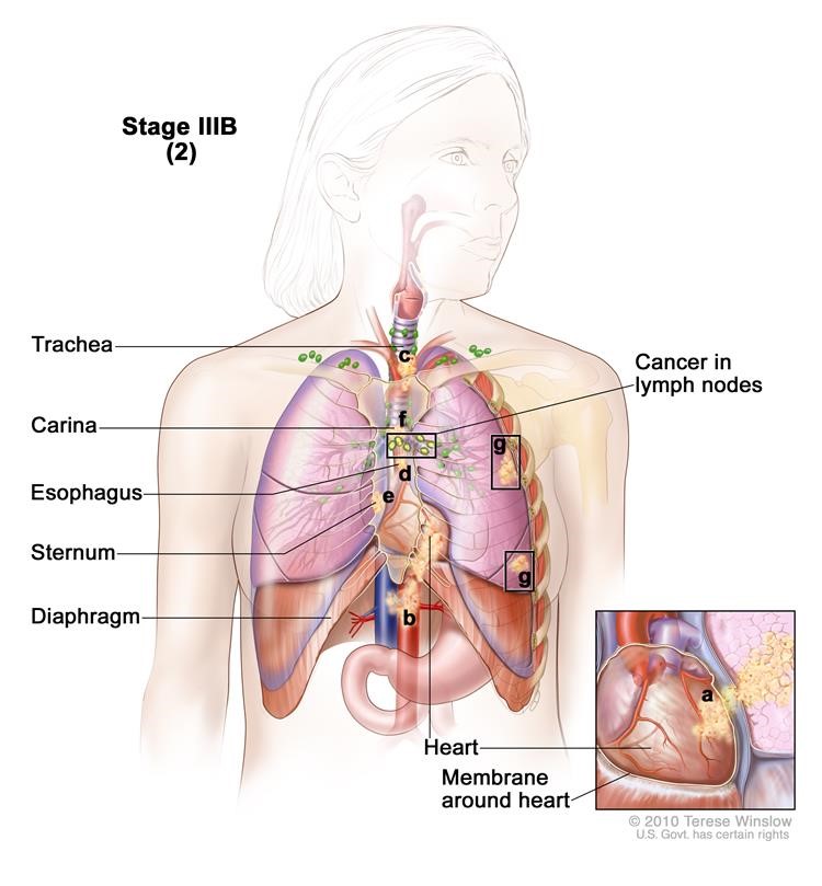
Figure 16: Stage IIIB non-small cell lung cancer.
Cancer has spread to lymph nodes on the same side of the chest as the tumor. The lymph nodes with cancer are near the sternum (chest bone) or where the bronchus enters the lung. Also:
- The tumor may be any size.
- There may be separate tumors in different lobes of the same lung.
- Cancer has spread to any of the following:
- Heart.
- Major blood vessels that lead to or from the heart.
- Trachea.
- Esophagus.
- Nerve that controls the larynx (voice box).
- Sternum (chest bone) or backbone.
- Carina (where the trachea joins the bronchi).
Stage IV
The cancer has spread to the other lung, and/or to lymph nodes, fluid around the lungs or heart, and/or other parts of the body, such as the brain, liver, adrenal gland, kidney, or bone.
In stage IV, the tumor may be any size and cancer may have spread to lymph nodes. One or more of the following is true:
- There are one or more tumors in both lungs.
- Cancer is found in fluid around the lungs or the heart.
- Cancer has spread to other parts of the body, such as the brain, liver, adrenal glands, kidneys, or bone.
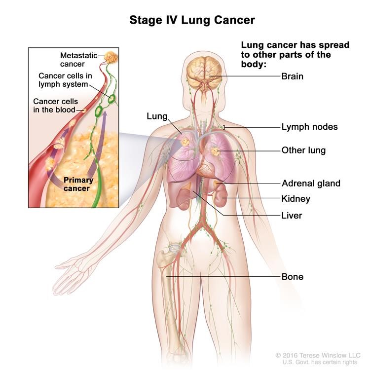
Figure 17: Stage IV non-small cell lung cancer.
Meeting With Your Surgeon
After these tests are completed, the thoracic surgeon will have a discussion with you about whether surgical repair is recommended.
If both you and the thoracic surgeon agree to proceed with surgery, the operation date will be booked.
This is a great opportunity to ask as many questions as you want.
Treatment Options Overview
There are different types of treatment for patients with non-small cell lung cancer.
Nine types of standard treatment are used:
- Surgery
- Radiation therapy
- Chemotherapy
- Targeted therapy
- Laser therapy
- Photodynamic therapy (PDT)
- Cryosurgery
- Electrocautery
- Watchful waiting
New types of treatment are being tested in clinical trials.
- Chemoprevention
- Radiosensitizers
- New combinations
Patients may want to think about taking part in a clinical trial.
Patients can enter clinical trials before, during, or after starting their cancer treatment.
Follow-up tests may be needed.
There are different types of treatment for patients with non-small cell lung cancer.
Different types of treatments are available for patients with non-small cell lung cancer. Some treatments are standard (the currently used treatment), and some are being tested in clinical trials. A treatment clinical trial is a research study meant to help improve current treatments or obtain information on new treatments for patients with cancer. When clinical trials show that a new treatment is better than the standard treatment, the new treatment may become the standard treatment. Patients may want to think about taking part in a clinical trial. Some clinical trials are open only to patients who have not started treatment.
Nine types of standard treatment are used:
Surgery
Four types of surgery are used to treat lung cancer:
Wedge resection:Surgery to remove a tumor and some of the normal tissue around it. When a slightly larger amount of tissue is taken, it is called a segmental resection.
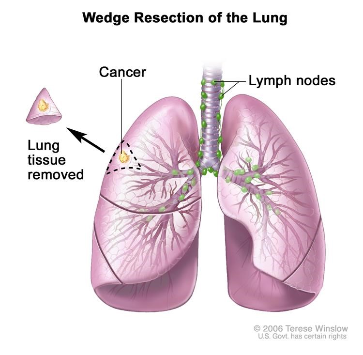
Figure 18: Wedge resection of the lung: Part of the lung lobe containing the cancer and a small amount of healthy tissue around it is removed.
Lobectomy: Surgery to remove a whole lobe (section) of the lung.
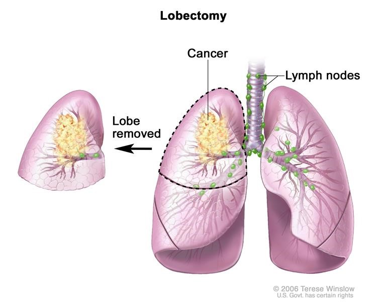
Figure 19: Lobectomy: A lobe of the lung is removed.
Pneumonectomy: Surgery to remove one whole lung.
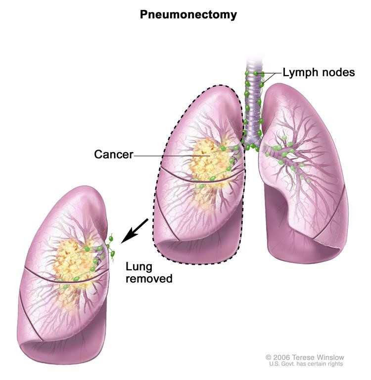
Figure 20: Pneumonectomy: The whole lung is removed.
Sleeve resection: Surgery to remove part of the bronchus.
Even if the doctor removes all the cancer that can be seen at the time of the surgery, some patients may be given chemotherapy or radiation therapy after surgery to kill any cancer cells that are left. Treatment given after the surgery, to lower the risk that the cancer will come back, is called adjuvant therapy.
Radiation therapy
Radiation therapy is a cancer treatment that uses high-energy x-rays or other types of radiation to kill cancer cells or keep them from growing. There are two types of radiation therapy:
- External radiation therapy uses a machine outside the body to send radiation toward the cancer.
- Internal radiation therapy uses a radioactive substance sealed in needles, seeds, wires, or catheters that are placed directly into or near the cancer.
Stereotactic body radiation therapy is a type of external radiation therapy. Special equipment is used to place the patient in the same position for each radiation treatment. Once a day for several days, a radiation machine aims a larger than usual dose of radiation directly at the tumor. By having the patient in the same position for each treatment, there is less damage to nearby healthy tissue. This procedure is also called stereotactic external-beam radiation therapy and stereotaxic radiation therapy.
Stereotactic radiosurgery is a type of external radiation therapy used to treat lung cancer that has spread to the brain. A rigid head frame is attached to the skull to keep the head still during the radiation treatment. A machine aims a single large dose of radiation directly at the tumor in the brain. This procedure does not involve surgery. It is also called stereotaxic radiosurgery, radiosurgery, and radiation surgery.
For tumors in the airways, radiation is given directly to the tumor through an endoscope.The way the radiation therapy is given depends on the type and stage of the cancer being treated. It also depends on where the cancer is found. External and internal radiation therapy are used to treat non-small cell lung cancer.
Chemotherapy
Chemotherapy is a cancer treatment that uses drugs to stop the growth of cancer cells, either by killing the cells or by stopping them from dividing. When chemotherapy is taken by mouth or injected into a vein or muscle, the drugs enter the bloodstream and can reach cancer cells throughout the body (systemic chemotherapy). When chemotherapy is placed directly into the cerebrospinal fluid, an organ, or a body cavity such as the abdomen, the drugs mainly affect cancer cells in those areas (regional chemotherapy).
The way the chemotherapy is given depends on the type and stage of the cancer being treated.
Targeted therapy
Targeted therapy is a type of treatment that uses drugs or other substances to attack specific cancer cells. Targeted therapies usually cause less harm to normal cells than chemotherapy or radiation therapy do. Monoclonal antibodies and tyrosine kinase inhibitors are the two main types of targeted therapy being used to treat advanced, metastatic, or recurrent non-small cell lung cancer.
Monoclonal antibodies
Monoclonal antibody therapy is a cancer treatment that uses antibodies made in the laboratory from a single type of immune system cell. These antibodies can identify substances on cancer cells or normal substances in the blood or tissues that may help cancer cells grow. The antibodies attach to the substances and kill the cancer cells, block their growth, or keep them from spreading. Monoclonal antibodies are given by infusion. They may be used alone or to carry drugs, toxins, or radioactive material directly to cancer cells.
There are different types of monoclonal antibody therapy:
Vascular endothelial growth factor (VEGF) inhibitor therapy: Cancer cells make a substance called VEGF, which causes new blood vessels to form (angiogenesis) and helps the cancer grow. VEGF inhibitors block VEGF and stop new blood vessels from forming. This may kill cancer cells because they need new blood vessels to grow. Bevacizumab and ramucirumab are VEGF inhibitors and angiogenesis inhibitors.
Epidermal growth factor receptor (EGFR) inhibitor therapy: EGFRs are proteins found on the surface of certain cells, including cancer cells. Epidermal growth factor attaches to the EGFR on the surface of the cell and causes the cells to grow and divide. EGFR inhibitors block the receptor and stop the epidermal growth factor from attaching to the cancer cell. This stops the cancer cell from growing and dividing. Cetuximab and necitumumab are EGFR inhibitors.
Immune checkpoint inhibitor therapy: PD-1 is a protein on the surface of T cells that helps keep the body’s immune responses in check. When PD-1 attaches to another protein called PDL-1 on a cancer cell, it stops the T cell from killing the cancer cell. PD-1 inhibitors attach to PDL-1 and allow the T cells to kill cancer cells. Nivolumab, pembrolizumab, and atezolizumab are types of immune checkpoint inhibitors.
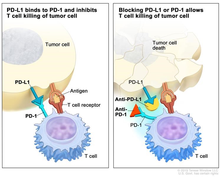
Figure 21: Immune checkpoint inhibitor: Checkpoint proteins, such as PD-L1 on tumor cells and PD-1 on T cells, help keep immune responses in check. The binding of PD-L1 to PD-1 keeps T cells from killing tumor cells in the body (left panel). Blocking the binding of PD-L1 to PD-1 with an immune checkpoint inhibitor (anti-PD-L1 or anti-PD-1) allows the T cells to kill tumor cells (right panel).
Tyrosine kinase inhibitors
Tyrosine kinase inhibitors are small-molecule drugs that go through the cell membrane and work inside cancer cells to block signals that cancer cells need to grow and divide. Some tyrosine kinase inhibitors also have angiogenesis inhibitor effects.
There are different types of tyrosine kinase inhibitors:
Epidermal growth factor receptor (EGFR) tyrosine kinase inhibitors: EGFRs are proteins found on the surface and inside certain cells, including cancer cells. Epidermal growth factor attaches to the EGFR inside the cell and sends signals to the tyrosine kinase area of the cell, which tells the cell to grow and divide. EGFR tyrosine kinase inhibitors stop these signals and stop the cancer cell from growing and dividing. Erlotinib, gefitinib, and afatinib are types of EGFR tyrosine kinase inhibitors. Some of these drugs work better when there is also a mutation (change) in the EGFR gene.
Kinase inhibitors that affect cells with certain gene changes: Certain changes in the ALK and ROS1 genes cause too much protein to be made. Blocking these proteins may stop the cancer from growing and spreading. Crizotinib is used to stop proteins from being made by the ALK and ROS1 gene. Ceritinib is used to stop proteins from being made by the ALK gene.
Staging Stage Specific Treatment of Lung Cancer
Stage I Non-Small Cell Lung Cancer
Treatment of stage I non-small cell lung cancer usually includes the following:
- Surgery (wedge resection, segmental resection, sleeve resection, or lobectomy).
- External radiation therapy, including stereotactic body radiation therapy for patients who cannot have surgery or choose not to have surgery.
Stage II Non-Small Cell Lung Cancer
Treatment of stage II non-small cell lung cancer may include the following:
- Surgery (wedge resection, segmental resection, sleeve resection, lobectomy, or pneumonectomy).
- Chemotherapy followed by surgery.
- Surgery followed by chemotherapy.
- External radiation therapy for patients who cannot have surgery.
Stage IIIA Non-Small Cell Lung Cancer
Treatment of stage IIIA non-small cell lung cancer that can be removed with surgery may include the following:
- Surgery followed by chemotherapy.
- Surgery followed by radiation therapy.
- Chemotherapy followed by surgery.
- Surgery followed by chemotherapy combined with radiation therapy.
- Chemotherapy and radiation therapy followed by surgery.
Stage IIIB Non-Small Cell Lung Cancer
Treatment of stage IIIB non-small cell lung cancer may include the following:
- Chemotherapy followed by external radiation therapy.
- Chemotherapy and radiation therapy given as separate treatments over the same period of time.
- External radiation therapy alone for patients who cannot be treated with chemotherapy.
- External radiation therapy as palliative therapy, to relieve symptoms and improve the quality of life.
Stage IV Non-Small Cell Lung Cancer
Treatment of stage IV non-small cell lung cancer may include the following:
- Chemotherapy.
- Chemotherapy followed by more chemotherapy as maintenance therapy to help keep cancer from progressing.
- Combination chemotherapy and targeted therapy with a monoclonal antibody, such as bevacizumab, cetuximab, or necitumumab.
- Targeted therapy alone.
- Targeted therapy with a tyrosine kinase inhibitor, such as erlotinib, gefitinib, afatinib, crizotinib, or ceritinib.
- External radiation therapy as palliative therapy, to relieve symptoms and improve the quality of life.
After Your Surgery (Post-Op)
How Long Will I Stay in Hospital?
How Will I Feel Post-Op?
Normal Symptoms After :
Contact Your Surgeon If:
What Kind of Activity Can I Do?
At the Hospital
The evening of your surgery, we will have you sitting in a chair and walking in the hallway. Being active reduces the risk of blood clots, and improves your breathing to prevent pneumonia.
The nursing staff on the surgical ward will insist on getting you out of bed!
At Home
While you should avoid strenuous activities during your recover, specific activities are recommended to promote a complete return to health and mobility.
Below are instructions on activity post-op:
Household Activities:
|
Driving: Avoid driving until you no longer need narcotic pain medication and you feel you can make rapid movements unimpaired by pain. Driving while taking narcotics can impair your ability to drive safely. |
|
|
Work: You should be able to return to work in X days. (how many days?) |
|
|
Sports:We suggest avoiding sports for 4 weeks. You can go for brisk walks 10 days after surgery. Wait 2 weeks before you swim. |
Taking Care of Your Incisions
When Can I Remove the Dressings?
A small white dressing is typically placed over each small incision from your minimally invasive lobectomy. These dressings may be removed 48 hours (2 days) after the operation.
Below this dressing are small brown or white pieces of tape (called Steri-Strips™); these will fall off on their own within 1-2 weeks. If they don’t fall off, you can remove them in the shower after two weeks.
If your surgeon gives you specific instructions other than this, please follow those instructions.
BathingYou can shower 48 hours after the surgery (after the gauze and clear tape dressing is removed). It is okay to get soap and water on the incisions. Pat the area dry. We recommend that you don’t submerge yourself in water (in a bathtub, pool, or hot-tub) for at least two weeks post-op. |
When to Contact Your Doctor
You should contact your doctor immediately if:
- You notice drainage from your incisions, particularly if it is thick and foul-smelling
- If you have a fever
- If you notice redness around your incisions, especially if it is getting worse
Pain Management and Medication
Although your surgery was likely completed using keyhole incisions you may have some pain. You will be given a PCA, a patient controlled analgesia, which allows you to press a button for additional medication when you need it.
At Osler we have an “Acute Pain Service” that is supervised by an anesthesiologist and a nurse practitioner. They will see you a few times to make sure you are comfortable
Your pain should be controlled well enough for you to begin getting out of bed the morning after surgery.
You will be discharged with pain medication, typically a mild to moderate strength narcotic. Take as needed and as prescribed.
Other Medications
You will be prescribed a stool softener when you go home. Narcotic or pain medications can cause constipation and a stool softener may help. If you have some nausea at home, you can pick up some Gravol® from the pharmacy.
Your Regular Medications
Unless otherwise instructed, continue to take your usual home medications once you are home.
Follow-Up
Unless otherwise instructed, continue to take your usual home medications once you are home.You will be given a date and time to see your surgeon following surgery, usually in 3 weeks.
The follow-up appointment should have been arranged during the office visit. If not, please call 905-458-4520 to arrange an appointment.
This information is produced and provided by the National Cancer Institute (NCI). The information in this topic may have changed since it was written. For the most current information, contact the National Cancer Institute.