This information is produced and provided by the National Cancer Institute (NCI). The information in this topic may have changed since it was written. For the most current information, contact the National Cancer Institute via the Internet web site at http://cancer.gov or call 1-800-4-CANCER.
General Information About Esophageal Cancer KEY POINTS
- Esophageal cancer is a disease in which malignant (cancer) cells form in the tissues of the esophagus.
- Smoking, heavy alcohol use, and Barrett esophagus can increase the risk of esophageal cancer.
- Signs and symptoms of esophageal cancer are weight loss and painful or difficult swallowing.
- Tests that examine the esophagus are used to detect (find) and diagnose esophageal cancer.
- Certain factors affect prognosis (chance of recovery) and treatment options.
Overview
Esophageal cancer is a disease in which malignant (cancer) cells form in the tissues of the esophagus.
The esophagus is the hollow, muscular tube that moves food and liquid from the throat to the stomach. The wall of the esophagus is made up of several layers of tissue, including mucous membrane, muscle, and connective tissue. Esophageal cancer starts on the inside lining of the esophagus and spreads outward through the other layers as it grows.
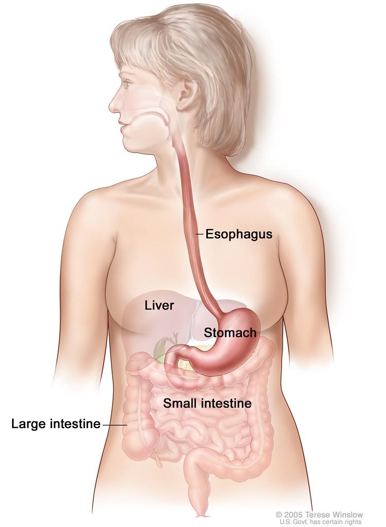
The esophagus and stomach are part of the upper gastrointestinal (digestive) system.
The two most common forms of esophageal cancer are named for the type of cells that become malignant (cancerous):
- Squamous cell carcinoma:Cancer that forms in squamous cells, the thin, flat cells lining the esophagus. This cancer is most often found in the upper and middle part of the esophagus, but can occur anywhere along the esophagus. This is also called epidermoid carcinoma.
- Adenocarcinoma: Cancer that begins inglandular (secretory) cells. Glandular cells in the lining of the esophagus produce and release fluids such as mucus. Adenocarcinomas usually form in the lower part of the esophagus, near the stomach.
Risk Factors
Smoking, heavy alcohol use, and Barrett esophagus can increase the risk of esophageal cancer.
Anything that increases your risk of getting a disease is called a risk factor. Having a risk factor does not mean that you will get cancer; not having risk factors doesn't mean that you will not get cancer. Talk with your doctor if you think you may be at risk.
Risk factors include the following:
- Tobacco use
- Heavy alcohol use
- Barrett esophagus: A condition in which the cells lining the lower part of the esophagus have changed or been replaced with abnormal cells that could lead to cancer of the esophagus. Gastric reflux (the backing up of stomach contents into the lower section of the esophagus) may irritate the esophagus and, over time, cause Barrett esophagus.
- Older age.
Signs and symptoms
Signs and symptoms of esophageal cancer are weight loss and painful or difficult swallowing.
These and other signs and symptoms may be caused by esophageal cancer or by other conditions. Check with your doctor if you have any of the following:
- Painful or difficult swallowing.
- Weight loss.
- Pain behind thebreastbone.
- Hoarseness and cough.
- Indigestion and heartburn.
Diagnose And Testing
Tests that examine the esophagus are used to detect (find) and diagnose esophageal cancer.
The following tests and procedures may be used:
-
Physical exam and history
An exam of the body to check general signs of health, including checking for signs of disease, such as lumps or anything else that seems unusual. A history of the patient’s health habits and past illnesses and treatments will also be taken. -
Chest x-ray
A x-ray of the organs and bones inside the chest. An x-ray is a type of energy beam that can go through the body and onto film, making a picture of areas inside the body. -
Barium swallow
A series of x-rays of the esophagus and stomach. The patient drinks a liquid that contains barium (a silver-white metallic compound). The liquid coats the esophagus and stomach, and x-rays are taken. This procedure is also called an upper GI series.
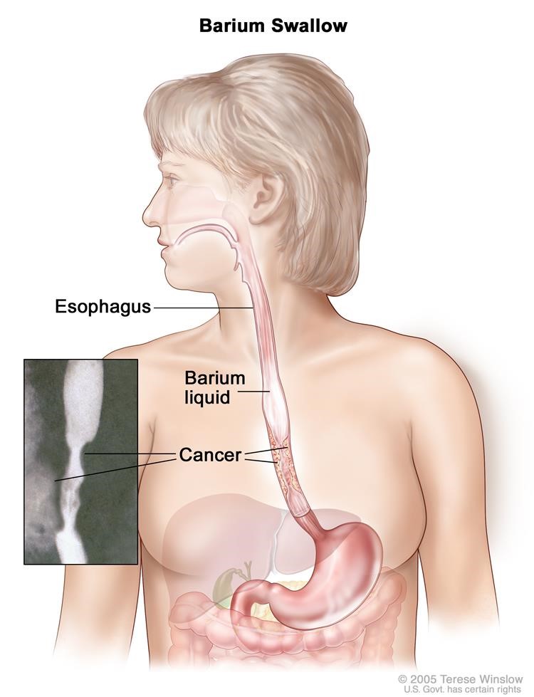
Barium swallow .The patient swallows barium liquid and it flows through the esophagus and into the stomach .X-rays are taken to look for abnormal areas.
-
Esophagoscopy
A procedure to look inside the esophagus to check for abnormal areas. An esophagoscope is inserted through the mouth or nose and down the throat into the esophagus. An esophagoscope is a thin, tube-like instrument with a light and a lens for viewing. It may also have a tool to remove tissue samples, which are checked under a microscope for signs of cancer. When the esophagus and stomach are looked at, it is called an upper endoscopy.
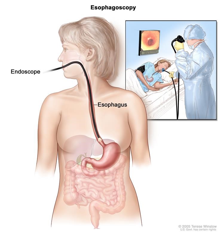
Esophagoscopy. A thin, lighted tube is inserted through the mouth and into the esophagus to look for abnormal areas.
-
Biopsy
The removal of cells or tissues so they can be viewed under a microscope by a pathologist to check for signs of cancer. The biopsy is usually done during an esophagoscopy. Sometimes a biopsy shows changes in the esophagus that are not cancer but may lead to cancer.
Certain factors affect prognosis (chance of recovery) and treatment options.
The prognosis (chance of recovery) and treatment options depend on the following:
- Thestage of the cancer (whether it affects part of the esophagus, involves the whole esophagus, or has spread to other places in the body).
- Whether the tumor can be completely removed bysurgery.
- The patient’s general health.
When esophageal cancer is found very early, there is a better chance of recovery. Esophageal cancer is often in an advanced stage when it is diagnosed. At later stages, esophageal cancer can be treated but rarely can be cured. Taking part in one of the clinical trials being done to improve treatment should be considered. Information about ongoing clinical trials is available from the NCI website.
Staging of esophageal cancer
KEY POINTS
- After esophageal cancer has been diagnosed, tests are done to find out if cancer cells have spread within the esophagus or to other parts of the body.
- There are three ways that cancer spreads in the body.
- Cancer may spread from where it began to other parts of the body.
- The grade of the tumor is also used to describe the cancer and plan treatment.
- The following stages are used for squamous cell carcinoma of the esophagus:
- Stage 0 (High-grade Dysplasia)
- Stage I squamous cell carcinoma of the esophagus
- Stage II squamous cell carcinoma of the esophagus
- Stage III squamous cell carcinoma of the esophagus
- Stage IV squamous cell carcinoma of the esophagus
- The following stages are used for adenocarcinoma of the esophagus:
- Stage 0 (High-grade Dysplasia)
- Stage I adenocarcinoma of the esophagus
- Stage II adenocarcinoma of the esophagus
- Stage III adenocarcinoma of the esophagus
- Stage IV adenocarcinoma of the esophagus
After esophageal cancer has been diagnosed, tests are done to find out if cancer cells have spread within the esophagus or to other parts of the body.
The process used to find out if cancer cells have spread within the esophagus or to other parts of the body is called staging. The information gathered from the staging process determines the stage of the disease. It is important to know the stage in order to plan treatment. The following tests and procedures may be used in the staging process:
-
Endoscopic ultrasound(EUS)
A procedure in which an endoscope is inserted into the body, usually through the mouth or rectum. For esophageal cancer, the endoscope is inserted through the mouth. An endoscope is a thin, tube-like instrument with a light and a lens for viewing. A probe at the end of the endoscope is used to bounce high-energy sound waves (ultrasound) off internal tissues or organs and make echoes. The echoes form a picture of body tissues called a sonogram. This procedure is also called endosonography. -
CT scan(CAT scan)
A procedure that makes a series of detailed pictures of areas inside the body, such as the chest, abdomen, and pelvis, taken from different angles. The pictures are made by a computer linked to an x-ray A dye may be injectedinto a vein or swallowed to help the organs or tissues show up more clearly. This procedure is also called computed tomography, computerized tomography, or computerized axial tomography. -
PET scan(positron emission tomography scan)
A procedure to find malignant tumorcells in the body. A small amount of radioactive glucose (sugar) is injected into a vein. The PET scanner rotates around the body and makes a picture of where glucose is being used in the body. Malignant tumor cells show up brighter in the picture because they are more active and take up more glucose than normal cells do. A PET scan and CT scan may be done at the same time. This is called a PET-CT. -
MRI(magnetic resonance imaging)
A procedure that uses a magnet, radio waves, and a computer to make a series of detailed pictures of areas inside the body. This procedure is also called nuclear magnetic resonance imaging (NMRI). -
Thoracoscopy
A surgical procedure to look at the organs inside the chest to check for abnormal areas. An incision (cut) is made between two ribs and a thoracoscope is inserted into the chest. A thoracoscope is a thin, tube-like instrument with a light and a lens for viewing. It may also have a tool to remove tissue or lymph node samples, which are checked under a microscope for signs of cancer. In some cases, this procedure may be used to remove part of the esophagus or lung. -
Laparoscopy
A surgical procedure to look at the organs inside the abdomen to check for signs of disease. Small incisions (cuts) are made in the wall of the abdomen and a laparoscope (a thin, lighted tube) is inserted into one of the incisions. Other instruments may be inserted through the same or other incisions to perform procedures such as removing organs or taking tissue samples to be checked under a microscope for signs of disease.
There are three ways that cancer spreads in the body.
Cancer can spread through tissue, the lymph system, and the blood:
- The cancer spreads from where it began by growing into nearby areas.
- Lymph system. The cancer spreads from where it began by getting into the lymph system. The cancer travels through thelymph vessels to other parts of the body.
- The cancer spreads from where it began by getting into the blood. The cancer travels through theblood vessels to other parts of the body.
Cancer may spread from where it began to other parts of the body.
When cancer spreads to another part of the body, it is called metastasis. Cancer cells break away from where they began (the primary tumor) and travel through the lymph system or blood.
- Lymph system. The cancer gets into the lymph system, travels through the lymph vessels, and forms atumor (metastatic tumor) in another part of the body.
- Blood. The cancer gets into the blood, travels through the blood vessels, and forms a tumor (metastatic tumor) in another part of the body.
The metastatic tumor is the same type of cancer as the primary tumor. For example, if esophageal cancer spreads to the lung, the cancer cells in the lung are actually esophageal cancer cells. The disease is metastatic esophageal cancer, not lung cancer.
The grade of the tumor is also used to describe the cancer and plan treatment.
The grade of the tumor describes how abnormal the cancer cells look under a microscope and how quickly the tumor is likely to grow and spread. Grades 1 to 3 are used to describe esophageal cancer:
- In grade 1, the cancer cells look more like normal cells under a microscope and grow and spread more slowly than grade 2 and 3 cancer cells.
- In grade 2, the cancer cells look more abnormal under a microscope and grow and spread more quickly than grade 1 cancer cells
- In grade 3, the cancer cells look more abnormal under a microscope and grow and spread more quickly than grade 1 and 2 cancer cells.
The following stages are used for squamous cell carcinoma of the esophagus:
Stage 0 (High-grade Dysplasia)
In stage 0, abnormal cells are found in the mucosa or submucosa layer of the esophaguswall. These abnormal cells may become cancer and spread into nearby normal tissue. Stage 0 is also called high-grade dysplasia.
Stage I squamous cell carcinoma of the esophagus
Stage I is divided into Stage IA and Stage IB, depending on where the cancer is found.
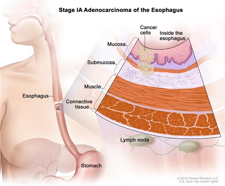
Stage IA: Cancer has formed in the mucosa or submucosa layer of the esophagus wall. The cancer cells are grade 1. Grade 1 cancer cells look more like normal cells under a microscope and grow and spread more slowly than grade 2 and 3 cancer cells.
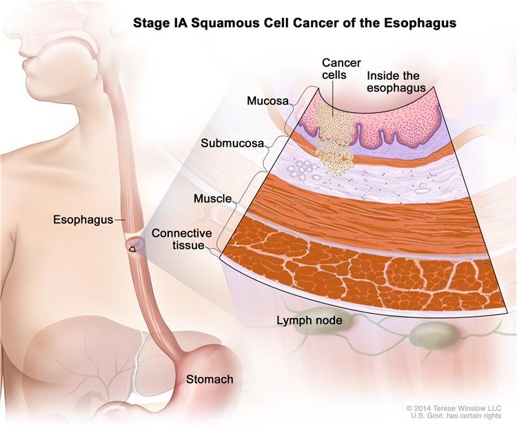
Stage IA squamous cell cancer of the esophagus.Cancer has formed in the mucosa or submucosa layer of the esophagus wall. The cancer cells are grade 1 or the grade is unknown. Grade 1 cancer cells look more like normal cells under a microscope and grow and spread more slowly than grade 2-3 cancer cells.
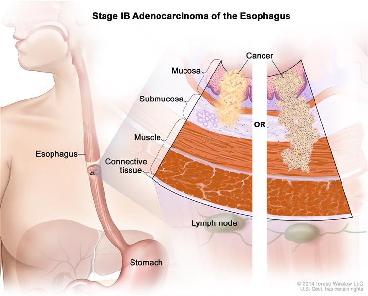
Stage IB: Cancer has formed:
- in the mucosaor submucosa layer of the esophagus The cancer cells are grade 2 and 3; or
- in the mucosa or submucosa layer and spread into the muscle layer or the connective tissuelayer of the esophagus wall. The cancer cells are grade 1. The tumor is in the lower esophagus or it is not known where the tumor is.
Grade 1 cancer cells look more like normal cells under a microscope and grow and spread more slowly than grade 2 and 3 cancer cells.
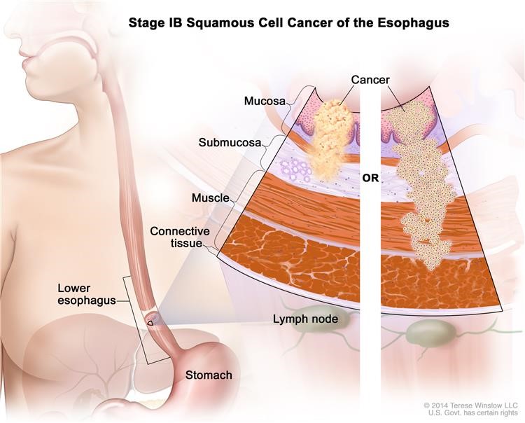
Stage IB squamous cell cancer of the esophagus. Cancer has formed in the mucosa or submucosa layer of the esophagus wall, and the cancer cells are grade 2-3; OR cancer has formed in the mucosa or submucosa layer and spread into the muscle layer or the connective tissue layer of the esophagus wall, and the cancer cells are grade 1. Grade 1 cancer cells look more like normal cells under a microscope and grow and spread more slowly than grade 2-3 cancer cells. The tumor is in the lower esophagus or it is not known where the tumor is.
Stage II squamous cell carcinoma of the esophagus
Stage II is divided into Stage IIA and Stage IIB, depending on where the cancer has spread.
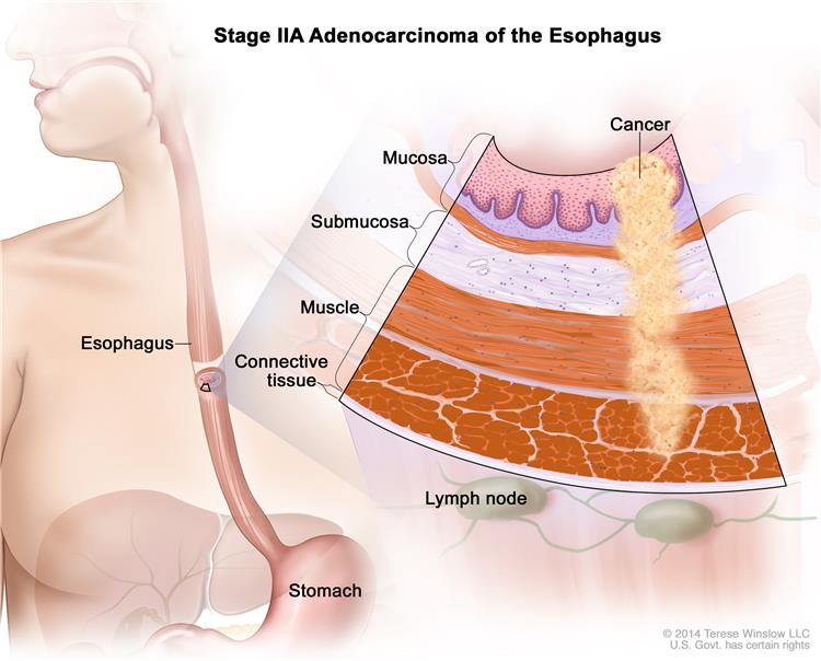
- Stage IIA: Cancer has spread:
- into the muscle layer or theconnective tissue layer of the esophagus The cancer cells are grade 1. The tumor is in either the upper or middle esophagus; or
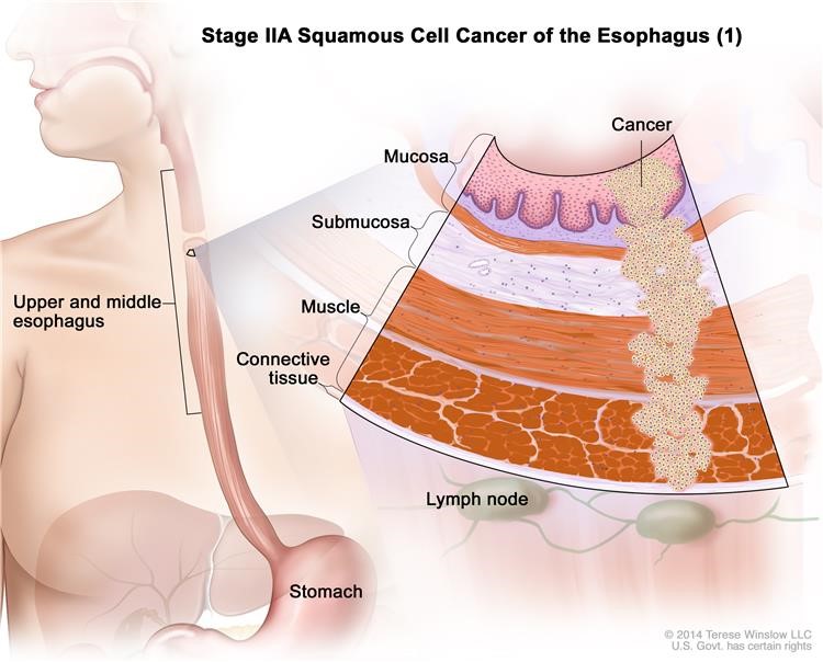
Stage IIA squamous cell cancer of the esophagus(1). The tumor is in either the upper or middle esophagus. Cancer has spread into the muscle layer or the connective tissue layer of the esophagus wall. The cancer cells are grade 1 . Grade 1 cancer cells look more like normal cells under a microscope and grow and spread more slowly than grade 2-3 cancer cells.
into the muscle layer or the connective tissue layer of the esophagus wall. The cancer cells are grade 2 and 3. The tumor is in the lower esophagus or it is not known where the tumor is.
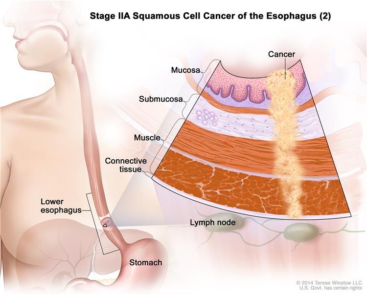
Stage IIA squamous cell cancer of the esophagus (2). The tumor is in the lower esophagus or it is not known where the tumor is. Cancer has spread into the muscle layer or the connective tissue layer of the esophagus wall. The cancer cells are grade 2-3 . Grade 2-3 cancer cells look more abnormal under a microscope and grow and spread more quickly than grade 1 cancer cells.
- Grade 1 cancer cells look more like normal cells under a microscopeand grow and spread more slowly than grade 2 and 3 cancer cells.
- Stage IIB: Cancer:
- has spread into the muscle layer or the connective tissuelayer of the esophagus The cancer cells are grade 2 and 3. Grade 2 and 3 cancer cells look more abnormal under a microscope and grow and spread more quickly than grade 1 cancer cells. The tumor is in either the upper or middle esophagus; or
- is in the mucosaor submucosa layer and may have spread into the muscle layer of the esophagus wall. Cancer is found in 1 or 2 lymph nodes near the tumor.
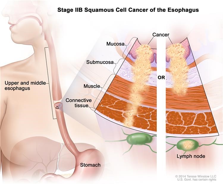
Stage IIB squamous cell cancer of the esophagus. The tumor is in either the upper or middle esophagus. Cancer has spread into the muscle layer or the connective tissue layer of the esophagus wall, and the cancer cells are grade 2-3; Or cancer is in the mucosa or submucosa layer and may have spread into the muscle layer of the esophagus wall, and cancer is found in 1 or 2 lymph nodes near the tumor. Grade 2-3 cancer cells look more abnormal under a microscope and grow and spread more quickly than grade 1 cancer cells.
Stage III squamous cell carcinoma of the esophagus
Stage III is divided into Stage IIIA, Stage IIIB, and Stage IIIC, depending on where the cancer has spread.
- Stage IIIA: Cancer:
- is in themucosa or submucosa layer and may have spread into the muscle layer of the esophagus Cancer is found in 3 to 6 lymph nodes near the tumor; or
- has spread into theconnective tissue layer of the esophagus wall. Cancer is found in 1 or 2 lymph nodes near the tumor; or
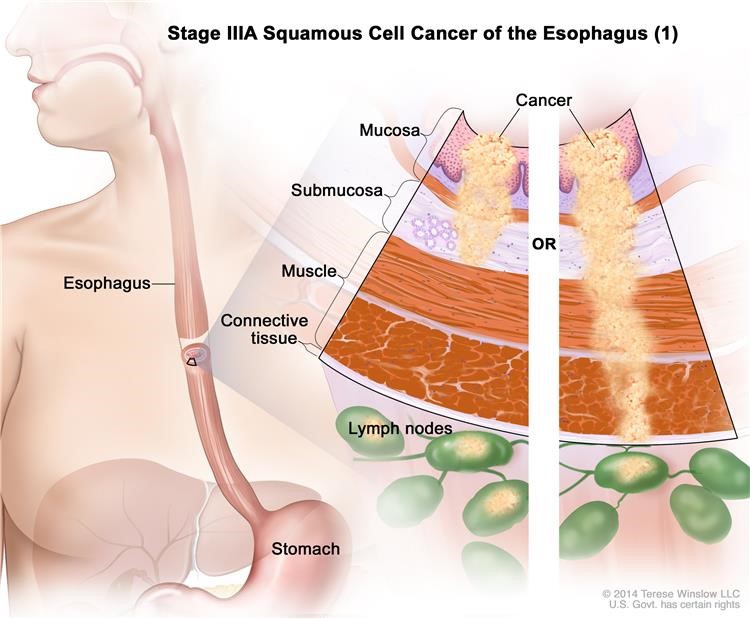
Stage IIIA squamous cell cancer of the esophagus (1). Cancer is in the mucosa or submucosa layer and may have spread into the muscle layer of the esophagus wall, and cancer is found in 3 to 6 lymph nodes near the tumor; OR cancer has spread into the connective tissue layer of the esophagus wall, and cancer is found in 1 or 2 lymph nodes near the tumor.
has spread into the diaphragm, pleura (tissue that covers the lungs and lines the inner wall of the chest cavity), or sac around the heart. The cancer can be removed by surgery.
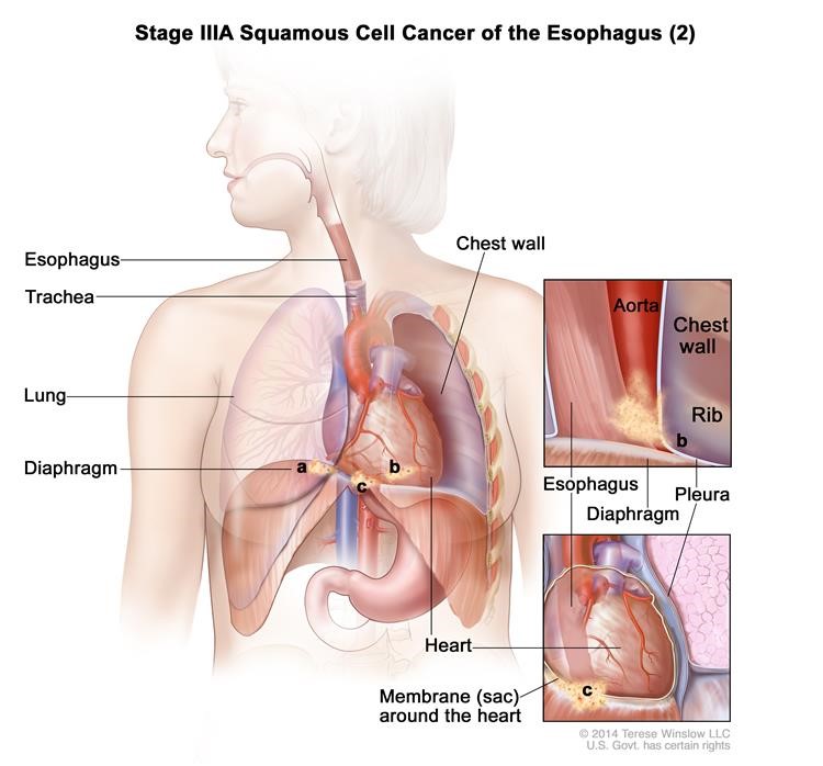
Stage IIIA squamous cell cancer of the esophagus (2). Cancer has spread into the (a) diaphragm, (b) pleura (tissue that covers the lungs and lines the inner wall of the chest cavity), or (c) membrane (sac) around the heart. The cancer can be removed by surgery.
Stage IIIB: Cancer has spread into the connective tissue layer of the esophagus wall. Cancer is found in 3 to 6 lymph nodes near the tumor.

Stage IIIB squamous cell cancer of the esophagus. Cancer has spread into the connective tissue layer of the esophagus wall. Cancer is found in 3 to 6 lymph nodes near the tumor.
Stage IIIC: Cancer has spread:
- into the diaphragm, pleura(tissue that covers the lungs and lines the inner wall of the chest cavity), or sac around the heart. The cancer can be removed by surgery. Cancer is found in 1 to 6 lymph nodes near the tumor; or
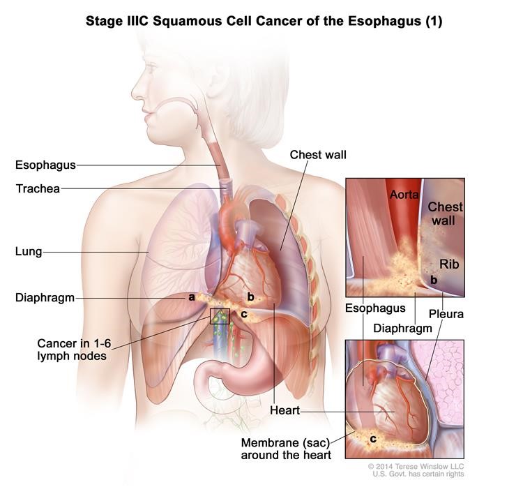
Stage IIIC squamous cell cancer of the esophagus (1). Cancer has spread into the (a) diaphragm, (b) pleura (tissue that covers the lungs and lines the inner wall of the chest cavity), or (c) membrane (sac) around the heart. The cancer can be removed by surgery. Cancer is found in 1 to 6 lymph nodes near the tumor.
- into other nearby organssuch as the aorta, trachea, or spine, and the cancer cannot be removed by surgery; or
- to 7 or more lymph nodes near the tumor.
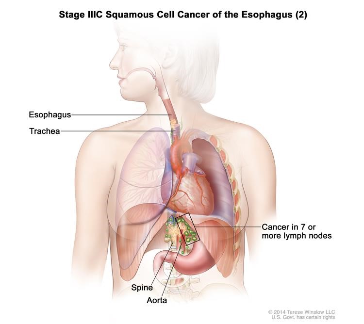
Stage IIIC squamous cell cancer of the esophagus (2). Cancer has spread into nearby organs, such as the aorta, trachea, or spine, and the cancer cannot be removed by surgery; OR cancer has spread to 7 or more lymph nodes near the tumor.
Stage IV squamous cell carcinoma of the esophagus
InStage IV,cancerhas spread to other parts of the body.
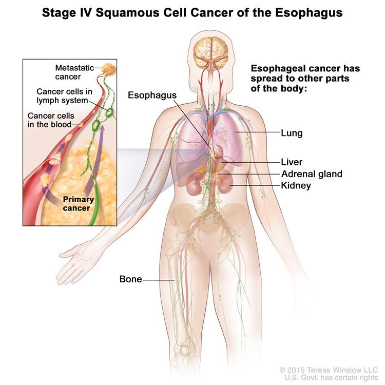
Stage IV squamous cell cancer of the esophagus. The cancer has spread to other parts of the body, such as the lung, liver, adrenal gland, kidney, or bone.
The following stages are used for adenocarcinoma of the esophagus:
Stage 0 (High-grade Dysplasia)
In stage 0, abnormal cells are found in the mucosa or submucosa layer of the esophaguswall. These abnormal cells may become cancer and spread into nearby normal tissue. Stage 0 is also called high-grade dysplasia.
Stage I adenocarcinoma of the esophagus
Stage I is divided into Stage IA and Stage IB, depending on where the cancer is found.
- Stage IA: Cancer has formed in themucosa or submucosa layer of the esophagus The cancer cells are grade 1 or 2. Grade 1 and 2 cancer cells look more like normal cells under a microscopeand grow and spread more slowly than grade 3 cancer cells.
Stage IA adenocarcinoma of the esophagus. Cancer has formed in the mucosa or submucosa layer of the esophagus wall. The Cancer cells are grade 1 or 2. Grade 1 and 2 cancer cells look more like normal cells under a microscope and grow and spread more slowly than grade 3 cancer cells.
Stage IB: Cancer has formed:
- in themucosaor submucosa layer of the esophagus The cancer cells are grade 3; or
- in the mucosa or submucosa layer and spread into the muscle layer of the esophagus wall. The cancer cells are grade 1 or 2.
Grade 1 and 2 cancer cells look more like normal cells under a microscope and grow and spread more slowly than grade 3 cancer cells.
Stage IB adenocarcinoma of the esophagus. Cancer has formed in the mucosa or submucosa layer of the esophagus wall, and the cancer cells are grade 3. Grade 3 cancer cells look more abnormal under a microscope and grow and spread more quickly than grade 1 or 2 cancer cells; OR cancer has formed in the mucosa or submucosa layer and spread into the muscle layer of the esophagus wall, and the cancer cells are grade 1 or 2. Grade 1 and 2 cancer cells look more like normal cells under a microscope and grow and spread more slowly than grade 3 cancer cells.
Stage II adenocarcinoma of the esophagus
Stage II is divided into Stage IIA and Stage IIB, depending on where the cancer has spread.
- Stage IIA: Cancer has spread into the muscle layer of theesophagus The cancer cells are grade 3. Grade 3 cancer cells look more abnormal under a microscope and grow and spread more quickly than grade 1 and 2 cancer cells.
Stage IIA adenocarcinoma of the esophagus. Cancer has spread into the muscle layer of the esophagus wall. The Cancer cells are grade 3. Grade 3 cancer cells look more abnormal under a microscope and grow and spread more quickly than grade 1 or 2 cancer cells.
Stage IIB: Cancer:
- has spread into the connective tissuelayer of the esophagus wall; or
- is in the mucosaor submucosa layer and may have spread into the muscle layer of the esophagus wall. Cancer is found in 1 or 2 lymph nodes near the tumor.
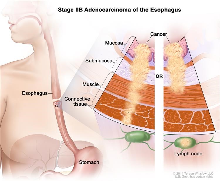
Stage IIB adenocarcinoma of the esophagus. Cancer has spread into the connective tissue layer of the esophagus wall; OR cancer is in the mucosa or submucosa layer and may have spread into the muscle layer of the esophagus wall, and cancer is found in 1 or 2 lymph nodes near the tumor.
Stage III adenocarcinoma of the esophagus
Stage III is divided into Stage IIIA, Stage IIIB, and Stage IIIC, depending on where the cancer has spread.
- Stage IIIA: Cancer:
- is in themucosa or submucosa layer and may have spread into the muscle layer of the esophagus Cancer is found in 3 to 6 lymph nodes near the tumor; or
has spread into the connective tissue layer of the esophagus wall. Cancer is found in 1 or 2 lymph nodes near the tumor; or
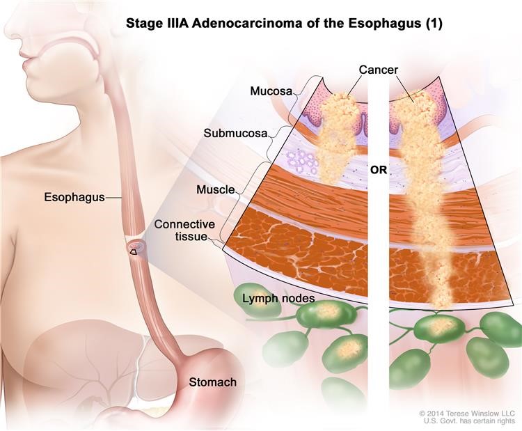
Stage IIIA adenocarcinoma of the esophagus (1). Cancer is in the mucosa or submucosa layer and may have spread into the muscle layer of the esophagus wall, and cancer is found in 3 to 6 lymph nodes near the tumor; OR cancer has spread into the connective tissue layer of the esophagus wall, cancer is found in 1 or 2 lymph nodes near the tumor.
has spread into the diaphragm, pleura (tissue that covers the lungs and lines the inner wall of the chest cavity), or sac around the heart. The cancer can be removed by surgery.
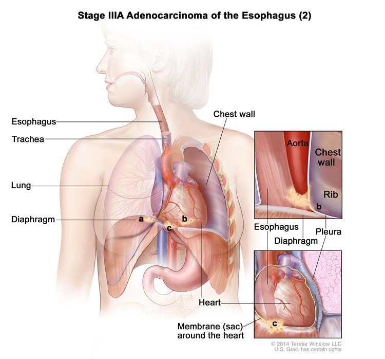
Stage IIIA adenocarcinoma of the esophagus (2). Cancer has spread into the (a) diaphragm, (b) pleura (tissue that covers the lungs and lines the inner wall of the chest cavity), or (c) membrane (sac) around the heart. The cancer can be removed by surgery.
Stage IIIB: Cancer has spread into the connective tissue layer of the esophagus wall. Cancer is found in 3 to 6 lymph nodes near the tumor.
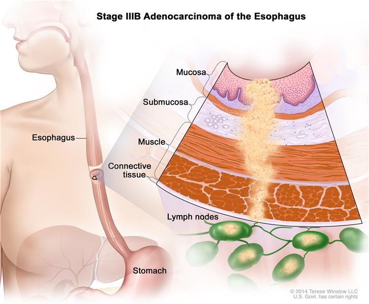
Stage IIIB adenocarcinoma of the esophagus. Cancer has spread into the connective tissue layer of the esophagus wall. Cancer is found in 3 to 6 lymph nodes near the tumor.
Stage IIIC: Cancer has spread:
- into the diaphragm, pleura(tissue that covers the lungs and lines the inner wall of the chest cavity), or sac around the heart. The cancer can be removed by surgery. Cancer is found in 1 to 6 lymph nodes near the tumor; or
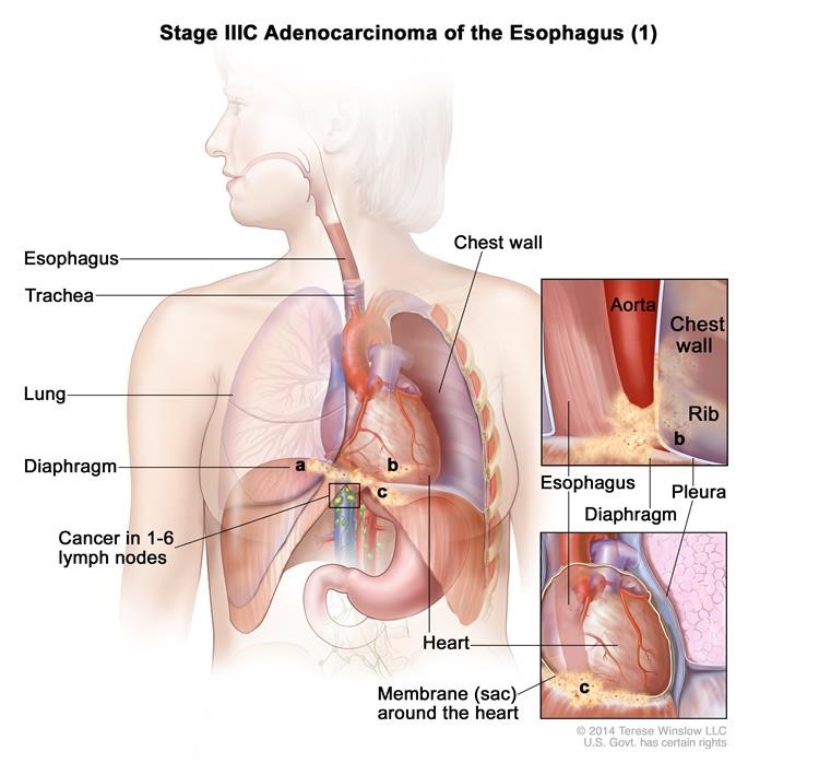
Stage IIIC adenocarcinoma of the esophagus (1). Cancer has spread into the (a) diaphragm, (b) pleura (tissue that covers the lungs and lines the inner wall of the chest cavity), or (c) membrane (sac) around the heart. The cancer can be removed by surgery. Cancer is found in 1 to 6 lymph nodes near the tumor.
- into other nearby organssuch as the aorta, trachea, or spine, and the cancer cannot be removed by surgery; or
- to 7 or more lymph nodes near the tumor.
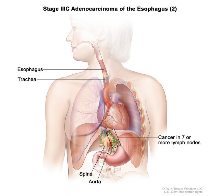
Stage IIIC adenocarcinoma of the esophagus (2). Cancer has spread into nearby organs, such as the aorta, trachea, or spine, and the cancer cannot be removed by surgery; OR cancer has spread to 7 or more lymph nodes near the tumor.
Stage IV adenocarcinoma of the esophagus
InStage IV, cancer has spread to other parts of the body.
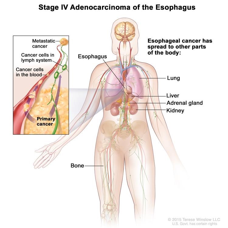
Stage IV adenocarcinoma of the esophagus. The cancer has spread to other parts of the body, such as the lung, liver, adrenal gland, kidney, or bone.
Recurrent Esophageal Cancer
Recurrent esophageal cancer is cancer that has recurred (come back) after it has been treated. The cancer may come back in the esophagusor in other parts of the body.
Treatment Options Overview
KEY POINTS
- There are different types of treatment for patients with esophageal cancer.
- Patients have special nutritional needs during treatment for esophageal cancer.
- Six types of standard treatment are used:
- Surgery
- Radiation therapy
- Chemotherapy
- Chemoradiation therapy
- Laser therapy
- Electrocoagulation
- New types of treatment are being tested in clinical trials.
- Targeted therapy
- Patients may want to think about taking part in a clinical trial.
- Patients can enter clinical trials before, during, or after starting their cancer treatment.
- Follow-up tests may be needed.
There are different types of treatment for patients with esophageal cancer.
Different types of treatment are available for patients with esophageal cancer. Some treatments are standard (the currently used treatment), and some are being tested in clinical trials.
A treatment clinical trial is a research study meant to help improve current treatments or obtain information on new treatments for patients with cancer. When clinical trials show that a new treatment is better than the standard treatment, the new treatment may become the standard treatment.
Patients may want to think about taking part in a clinical trial. Some clinical trials are open only to patients who have not started treatment.
Patients have special nutritional needs during treatment for esophageal cancer.
Many people with esophageal cancer find it hard to eat because they have trouble swallowing. The esophagus may be narrowed by the tumor or as a side effect of treatment. Some patients may receive nutrients directly into a vein. Others may need a feeding tube (a flexible plastic tube that is passed through the nose or mouth into the stomach) until they are able to eat on their own.
Six types of standard treatment are used:
Surgery
Surgery is the most common treatment for cancer of the esophagus. Part of the esophagus may be removed in an operation called an esophagectomy.
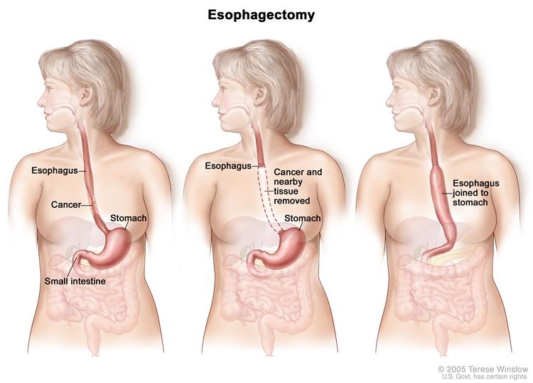
Esophagectomy. A portion of the esophagus is removed and the stomach is pulled up and joined to the remaining esophagus.
The doctor will connect the remaining healthy part of the esophagus to the stomach so the patient can still swallow. A plastic tube or part of the intestine may be used to make the connection. Lymph nodes near the esophagus may also be removed and viewed under a microscope to see if they contain cancer. If the esophagus is partly blocked by the tumor, an expandable metal stent (tube) may be placed inside the esophagus to help keep it open.
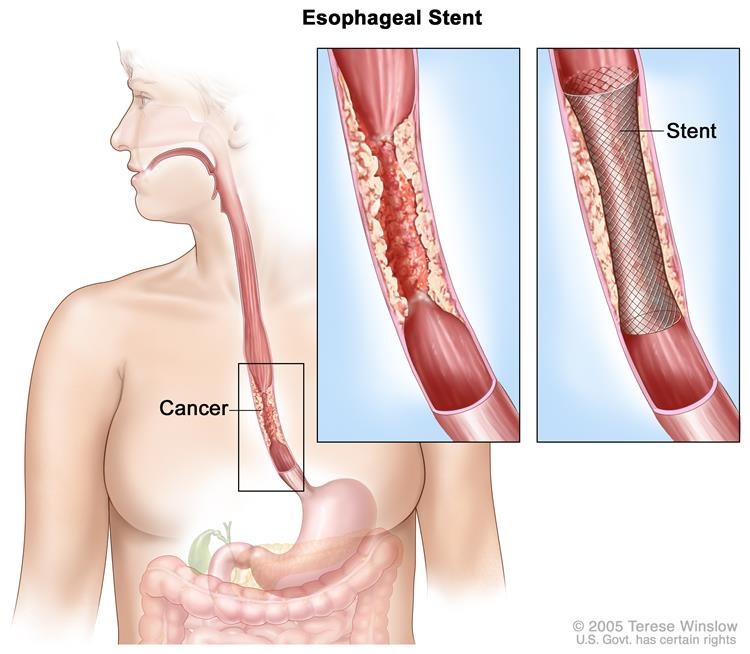
Esophageal stent. A device (stent) is placed in the esophagus to keep it open to allow food and liquids to pass through into the stomach.
Small,early-stage cancer and high-grade dysplasia of the esophagus may be removed by endoscopic resection. An endoscope (a thin, tube-like instrument with a light and a lens for viewing) is inserted through a small incision (cut) in the skin or through an opening in the body, such as the mouth. A tool attached to the endoscope is used to remove tissue.
Radiation therapy
Radiation therapy is a cancer treatment that uses high-energy x-rays or other types of radiation to kill cancer cells or keep them from growing. There are two types of radiation therapy:
- External radiation therapyuses a machine outside the body to send radiation toward the cancer.
- Internal radiation therapyuses a radioactive substance sealed in needles, seeds, wires, or catheters that are placed directly into or near the cancer.
The way the radiation therapy is given depends on the type and stage of the cancer being treated. External and internal radiation therapy are used to treat esophageal cancer.
A plastic tube may be inserted into the esophagus to keep it open during radiation therapy. This is called intraluminal intubation and dilation.
Chemotherapy
Chemotherapy is a cancer treatment that uses drugs to stop the growth of cancer cells, either by killing the cells or by stopping them from dividing. When chemotherapy is taken by mouth or injected into a vein or muscle, the drugs enter the bloodstream and can reach cancer cells throughout the body (systemic chemotherapy). When chemotherapy is placed directly into the cerebrospinal fluid, an organ, or a body cavity such as the abdomen, the drugs mainly affect cancer cells in those areas (regional chemotherapy). The way the chemotherapy is given depends on the type and stage of the cancer being treated.
See Drugs Approved for Esophageal Cancer for more information.
Chemoradiation therapy
Chemoradiation therapy combines chemotherapy and radiation therapy to increase the effects of both.
Laser therapy
Laser therapy is a cancer treatment that uses a laser beam (a narrow beam of intense light) to kill cancer cells.
Electrocoagulation
Electrocoagulation is the use of an electric current to kill cancer cells.
New types of treatment are being tested in clinical trials.
This summary section describes treatments that are being studied in clinical trials. It may not mention every new treatment being studied. Information about clinical trials is available from the NCI website.
Targeted therapy
Targeted therapy is a type of treatment that uses drugs or other substances to identify and attack specific cancer cells. Targeted therapies usually cause less harm to normal cells than chemotherapy or radiation therapy do. Monoclonal antibody therapy is a type of targeted therapy used in the treatment of esophageal cancer.
Monoclonal antibody therapy uses antibodies made in the laboratory from a single type of immune system cell. These antibodies can identify substances on cancer cells or normal substances that may help cancer cells grow. The antibodies attach to the substances and kill the cancer cells, block their growth, or keep them from spreading. Monoclonal antibodies are given by infusion. They may be used alone or to carry drugs, toxins, or radioactive material directly to cancer cells. Trastuzumab is a monoclonal antibody being studied in esophageal cancer. It may be given to block the effect of the growth factorprotein HER2, which sends growth signals to esophageal cancer cells.
Patients may want to think about taking part in a clinical trial.
For some patients, taking part in a clinical trial may be the best treatment choice. Clinical trials are part of the cancer research process. Clinical trials are done to find out if new cancer treatments are safe and effective or better than the standard treatment.
Many of today's standard treatments for cancer are based on earlier clinical trials. Patients who take part in a clinical trial may receive the standard treatment or be among the first to receive a new treatment.
Patients who take part in clinical trials also help improve the way cancer will be treated in the future. Even when clinical trials do not lead to effective new treatments, they often answer important questions and help move research forward.
Patients can enter clinical trials before, during, or after starting their cancer treatment.
Some clinical trials only include patients who have not yet received treatment. Other trials test treatments for patients whose cancer has not gotten better. There are also clinical trials that test new ways to stop cancer from recurring (coming back) or reduce the side effects of cancer treatment.
Clinical trials are taking place in many parts of the country. See the Treatment Options section that follows for links to current treatment clinical trials. These have been retrieved from NCI's listing of clinical trials.
Follow-up tests may be needed
Some of the tests that were done to diagnose the cancer or to find out the stage of the cancer may be repeated. Some tests will be repeated in order to see how well the treatment is working. Decisions about whether to continue, change, or stop treatment may be based on the results of these tests.
Some of the tests will continue to be done from time to time after treatment has ended. The results of these tests can show if your condition has changed or if the cancer has recurred (come back). These tests are sometimes called follow-uptests or check-ups.
Treatment Options By Stage
- Stage 0 (High-grade Dysplasia)
- Stage I Esophageal Cancer
- Stage II Esophageal Cancer
- Stage III Esophageal Cancer
- Stage IV Esophageal Cancer
Stage 0 (High-grade Dysplasia)
Treatment of stage 0 may include the following:
Check the list of NCI-supported cancer clinical trials that are now accepting patients with stage 0 esophageal cancer. For more specific results, refine the search by using other search features, such as the location of the trial, the type of treatment, or the name of the drug. Talk with your doctor about clinical trials that may be right for you. General information about clinical trials is available from the NCI website.
Stage I Esophageal Cancer
Treatment of stage I esophageal squamous cell carcinoma or adenocarcinoma may include the following:
- Chemoradiation therapyfollowed by surgery.
- Surgery alone.
Check the list of NCI-supported cancer clinical trials that are now accepting patients with stage I esophageal cancer. For more specific results, refine the search by using other search features, such as the location of the trial, the type of treatment, or the name of the drug. Talk with your doctor about clinical trials that may be right for you. General information about clinical trials is available from the NCI website.
Stage II Esophageal Cancer
Treatment of stage II esophageal squamous cell carcinoma or adenocarcinoma may include the following:
- Chemoradiation therapyfollowed by surgery.
- Surgery alone.
- Chemotherapyfollowed by surgery.
- Chemoradiation therapy alone.
Check the list of NCI-supported cancer clinical trials that are now accepting patients with stage II esophageal cancer. For more specific results, refine the search by using other search features, such as the location of the trial, the type of treatment, or the name of the drug. Talk with your doctor about clinical trials that may be right for you. General information about clinical trials is available from the NCI website.
Stage III Esophageal Cancer
Treatment of stage III esophageal squamous cell carcinoma or adenocarcinoma may include the following:
- Chemoradiation therapyfollowed by surgery.
- Chemotherapyfollowed by surgery.
- Chemoradiation therapy alone.
Check the list of NCI-supported cancer clinical trials that are now accepting patients with stage III esophageal cancer. For more specific results, refine the search by using other search features, such as the location of the trial, the type of treatment, or the name of the drug. Talk with your doctor about clinical trials that may be right for you. General information about clinical trials is available from the NCI website.
Stage IV Esophageal Cancer
Treatment of stage IV esophageal squamous cell carcinoma or adenocarcinoma may include the following:
- Chemoradiation therapyfollowed by surgery.
- Chemotherapy.
- Laser surgeryor electrocoagulation as palliative therapy to relieve symptoms and improve quality of life.
- Anesophageal stent as palliative therapy to relieve symptoms and improve quality of life.
- Externalor internal radiation therapy as palliative therapy to relieve symptoms and improve quality of life.
- Clinical trialsof chemotherapy.
- A clinical trial oftargeted therapy combined with chemotherapy.
Check the list of NCI-supported cancer clinical trials that are now accepting patients with stage IV esophageal cancer. For more specific results, refine the search by using other search features, such as the location of the trial, the type of treatment, or the name of the drug. Talk with your doctor about clinical trials that may be right for you. General information about clinical trials is available from the NCI website.
Treatment Options for Recurrent Esophageal Cancer
Treatment of recurrent esophageal cancer may include the following:
- Use of anystandard treatments as palliative therapy to relieve symptoms and improve quality of life.
- Clinical trials.
Check the list of NCI-supported cancer clinical trials that are now accepting patients with recurrent esophageal cancer. For more specific results, refine the search by using other search features, such as the location of the trial, the type of treatment, or the name of the drug. Talk with your doctor about clinical trials that may be right for you. General information about clinical trials is available from the NCI website.
To Learn More About Esophageal Cancer
For more information from the National Cancer Institute about esophageal cancer, see the following:
- Esophageal Cancer Home Page
- What You Need to Know About™ Cancer of the Esophagus
- Esophageal Cancer Prevention
- Esophageal Cancer Screening
- Unusual Cancers of Childhood Treatment
- Gastrointestinal Stromal Tumors Treatment
- Nutrition in Cancer Care
- Oral Complications of Chemotherapy and Head/Neck Radiation
- Tobacco(includes help with quitting)
- Lasers in Cancer Treatment
For general cancer information and other resources from the National Cancer Institute, see the following:
- About Cancer
- Staging
- Chemotherapy and You: Support for People With Cancer
- Radiation Therapy and You: Support for People With Cancer
- Coping with Cancer
- Questions to Ask Your Doctor about Cancer
- For Survivors and Caregivers
About This PDQ Summary
About PDQ
Physician Data Query (PDQ) is the National Cancer Institute's (NCI's) comprehensive cancer information database. The PDQ database contains summaries of the latest published information on cancer prevention, detection, genetics, treatment, supportive care, and complementary and alternative medicine. Most summaries come in two versions. The health professional versions have detailed information written in technical language. The patient versions are written in easy-to-understand, nontechnical language. Both versions have cancer information that is accurate and up to date and most versions are also available in Spanish.
PDQ is a service of the NCI. The NCI is part of the National Institutes of Health (NIH). NIH is the federal government’s center of biomedical research. The PDQ summaries are based on an independent review of the medical literature. They are not policy statements of the NCI or the NIH.
Purpose of This Summary
This PDQ cancer information summary has current information about the treatment of esophageal cancer. It is meant to inform and help patients, families, and caregivers. It does not give formal guidelines or recommendations for making decisions about health care.
Reviewers and Updates
Editorial Boards write the PDQ cancer information summaries and keep them up to date. These Boards are made up of experts in cancer treatment and other specialties related to cancer. The summaries are reviewed regularly and changes are made when there is new information. The date on each summary ("Date Last Modified") is the date of the most recent change.
The information in this patient summary was taken from the health professional version, which is reviewed regularly and updated as needed, by the PDQ Adult Treatment Editorial Board.
Clinical Trial Information
A clinical trial is a study to answer a scientific question, such as whether one treatment is better than another. Trials are based on past studies and what has been learned in the laboratory. Each trial answers certain scientific questions in order to find new and better ways to help cancer patients. During treatment clinical trials, information is collected about the effects of a new treatment and how well it works. If a clinical trial shows that a new treatment is better than one currently being used, the new treatment may become "standard." Patients may want to think about taking part in a clinical trial. Some clinical trials are open only to patients who have not started treatment.
Clinical trials are listed in PDQ and can be found online at NCI's website. Many cancer doctors who take part in clinical trials are also listed in PDQ. For more information, call the Cancer Information Service 1-800-4-CANCER (1-800-422-6237).
Disclaimer
The information in these summaries should not be used to make decisions about insurance reimbursement. More information on insurance coverage is available on Cancer.gov on the Managing Cancer Care page.